Related Publications
2576724
IXMHRBF6
image software
1
apa-cv
50
date
desc
1
1
4123
https://labsyspharm.org/wp-content/plugins/zotpress/
%7B%22status%22%3A%22success%22%2C%22updateneeded%22%3Afalse%2C%22instance%22%3Afalse%2C%22meta%22%3A%7B%22request_last%22%3A0%2C%22request_next%22%3A0%2C%22used_cache%22%3Atrue%7D%2C%22data%22%3A%5B%7B%22key%22%3A%22GWZ3DV7V%22%2C%22library%22%3A%7B%22id%22%3A2576724%7D%2C%22meta%22%3A%7B%22lastModifiedByUser%22%3A%7B%22id%22%3A6889081%2C%22username%22%3A%22arenasg%22%2C%22name%22%3A%22%22%2C%22links%22%3A%7B%22alternate%22%3A%7B%22href%22%3A%22https%3A%5C%2F%5C%2Fwww.zotero.org%5C%2Farenasg%22%2C%22type%22%3A%22text%5C%2Fhtml%22%7D%7D%7D%2C%22creatorSummary%22%3A%22de%20Bruijn%20et%20al.%22%2C%22parsedDate%22%3A%222025-04%22%2C%22numChildren%22%3A1%7D%2C%22bib%22%3A%22%3Cdiv%20class%3D%5C%22csl-bib-body%5C%22%20style%3D%5C%22line-height%3A%202%3B%20padding-left%3A%201em%3B%20text-indent%3A-1em%3B%5C%22%3E%5Cn%20%20%3Cdiv%20class%3D%5C%22csl-entry%5C%22%3Ede%20Bruijn%2C%20I.%2C%20Nikolov%2C%20M.%2C%20Lau%2C%20C.%2C%20Clayton%2C%20A.%2C%20Gibbs%2C%20D.%20L.%2C%20Mitraka%2C%20E.%2C%20Pozhidayeva%2C%20D.%2C%20Lash%2C%20A.%2C%20Sumer%2C%20S.%20O.%2C%20Altreuter%2C%20J.%2C%20Anton%2C%20K.%2C%20DeFelice%2C%20M.%2C%20Li%2C%20X.%2C%20Lisman%2C%20A.%2C%20Longabaugh%2C%20W.%20J.%20R.%2C%20Muhlich%2C%20J.%2C%20Santagata%2C%20S.%2C%20Nandakumar%2C%20S.%2C%20Sorger%2C%20P.%20K.%2C%20%26%23x2026%3B%20Eddy%2C%20J.%20A.%20%282025%29.%20Sharing%20data%20from%20the%20Human%20Tumor%20Atlas%20Network%20through%20standards%2C%20infrastructure%20and%20community%20engagement.%20%3Ci%3ENature%20Methods%3C%5C%2Fi%3E%2C%20%3Ci%3E22%3C%5C%2Fi%3E%284%29%2C%20664%26%23x2013%3B671.%20%3Ca%20class%3D%27zp-DOIURL%27%20target%3D%27_blank%27%20href%3D%27https%3A%5C%2F%5C%2Fdoi.org%5C%2F10.1038%5C%2Fs41592-025-02643-0%27%3Ehttps%3A%5C%2F%5C%2Fdoi.org%5C%2F10.1038%5C%2Fs41592-025-02643-0%3C%5C%2Fa%3E%3C%5C%2Fdiv%3E%5Cn%3C%5C%2Fdiv%3E%22%2C%22data%22%3A%7B%22itemType%22%3A%22journalArticle%22%2C%22title%22%3A%22Sharing%20data%20from%20the%20Human%20Tumor%20Atlas%20Network%20through%20standards%2C%20infrastructure%20and%20community%20engagement%22%2C%22creators%22%3A%5B%7B%22creatorType%22%3A%22author%22%2C%22firstName%22%3A%22Ino%22%2C%22lastName%22%3A%22de%20Bruijn%22%7D%2C%7B%22creatorType%22%3A%22author%22%2C%22firstName%22%3A%22Milen%22%2C%22lastName%22%3A%22Nikolov%22%7D%2C%7B%22creatorType%22%3A%22author%22%2C%22firstName%22%3A%22Clarisse%22%2C%22lastName%22%3A%22Lau%22%7D%2C%7B%22creatorType%22%3A%22author%22%2C%22firstName%22%3A%22Ashley%22%2C%22lastName%22%3A%22Clayton%22%7D%2C%7B%22creatorType%22%3A%22author%22%2C%22firstName%22%3A%22David%20L.%22%2C%22lastName%22%3A%22Gibbs%22%7D%2C%7B%22creatorType%22%3A%22author%22%2C%22firstName%22%3A%22Elvira%22%2C%22lastName%22%3A%22Mitraka%22%7D%2C%7B%22creatorType%22%3A%22author%22%2C%22firstName%22%3A%22Dar%27ya%22%2C%22lastName%22%3A%22Pozhidayeva%22%7D%2C%7B%22creatorType%22%3A%22author%22%2C%22firstName%22%3A%22Alex%22%2C%22lastName%22%3A%22Lash%22%7D%2C%7B%22creatorType%22%3A%22author%22%2C%22firstName%22%3A%22Selcuk%20Onur%22%2C%22lastName%22%3A%22Sumer%22%7D%2C%7B%22creatorType%22%3A%22author%22%2C%22firstName%22%3A%22Jennifer%22%2C%22lastName%22%3A%22Altreuter%22%7D%2C%7B%22creatorType%22%3A%22author%22%2C%22firstName%22%3A%22Kristen%22%2C%22lastName%22%3A%22Anton%22%7D%2C%7B%22creatorType%22%3A%22author%22%2C%22firstName%22%3A%22Mialy%22%2C%22lastName%22%3A%22DeFelice%22%7D%2C%7B%22creatorType%22%3A%22author%22%2C%22firstName%22%3A%22Xiang%22%2C%22lastName%22%3A%22Li%22%7D%2C%7B%22creatorType%22%3A%22author%22%2C%22firstName%22%3A%22Aaron%22%2C%22lastName%22%3A%22Lisman%22%7D%2C%7B%22creatorType%22%3A%22author%22%2C%22firstName%22%3A%22William%20J.%20R.%22%2C%22lastName%22%3A%22Longabaugh%22%7D%2C%7B%22creatorType%22%3A%22author%22%2C%22firstName%22%3A%22Jeremy%22%2C%22lastName%22%3A%22Muhlich%22%7D%2C%7B%22creatorType%22%3A%22author%22%2C%22firstName%22%3A%22Sandro%22%2C%22lastName%22%3A%22Santagata%22%7D%2C%7B%22creatorType%22%3A%22author%22%2C%22firstName%22%3A%22Subhiksha%22%2C%22lastName%22%3A%22Nandakumar%22%7D%2C%7B%22creatorType%22%3A%22author%22%2C%22firstName%22%3A%22Peter%20K.%22%2C%22lastName%22%3A%22Sorger%22%7D%2C%7B%22creatorType%22%3A%22author%22%2C%22firstName%22%3A%22Christine%22%2C%22lastName%22%3A%22Suver%22%7D%2C%7B%22creatorType%22%3A%22author%22%2C%22firstName%22%3A%22Xengie%22%2C%22lastName%22%3A%22Doan%22%7D%2C%7B%22creatorType%22%3A%22author%22%2C%22firstName%22%3A%22Justin%22%2C%22lastName%22%3A%22Guinney%22%7D%2C%7B%22creatorType%22%3A%22author%22%2C%22firstName%22%3A%22Nikolaus%22%2C%22lastName%22%3A%22Schultz%22%7D%2C%7B%22creatorType%22%3A%22author%22%2C%22firstName%22%3A%22Adam%20J.%22%2C%22lastName%22%3A%22Taylor%22%7D%2C%7B%22creatorType%22%3A%22author%22%2C%22firstName%22%3A%22V%5Cu00e9steinn%22%2C%22lastName%22%3A%22Thorsson%22%7D%2C%7B%22creatorType%22%3A%22author%22%2C%22firstName%22%3A%22Ethan%22%2C%22lastName%22%3A%22Cerami%22%7D%2C%7B%22creatorType%22%3A%22author%22%2C%22firstName%22%3A%22James%20A.%22%2C%22lastName%22%3A%22Eddy%22%7D%5D%2C%22abstractNote%22%3A%22Data%20from%20the%20first%20phase%20of%20the%20Human%20Tumor%20Atlas%20Network%20%28HTAN%29%20are%20now%20available%2C%20comprising%208%2C425%20biospecimens%20from%202%2C042%20research%20participants%20profiled%20with%20more%20than%2020%20molecular%20assays.%20The%20data%20were%20generated%20to%20study%20the%20evolution%20from%20precancerous%20to%20advanced%20disease.%20The%20HTAN%20Data%20Coordinating%20Center%20%28DCC%29%20has%20enabled%20their%20dissemination%20and%20effective%20reuse.%20We%20describe%20the%20diverse%20datasets%2C%20how%20to%20access%20them%2C%20data%20standards%2C%20underlying%20infrastructure%20and%20governance%20approaches%2C%20and%20our%20methods%20to%20sustain%20community%20engagement.%20HTAN%20data%20can%20be%20accessed%20through%20the%20HTAN%20Portal%2C%20explored%20in%20visualization%20tools-including%20CellxGene%2C%20Minerva%20and%20cBioPortal-and%20analyzed%20in%20the%20cloud%20through%20the%20NCI%20Cancer%20Research%20Data%20Commons.%20Infrastructure%20was%20developed%20to%20enable%20data%20ingestion%20and%20dissemination%20through%20the%20Synapse%20platform.%20The%20HTAN%20DCC%27s%20flexible%20and%20modular%20approach%20to%20sharing%20complex%20cancer%20research%20data%20offers%20valuable%20insights%20to%20other%20data-coordination%20efforts%20and%20researchers%20looking%20to%20leverage%20HTAN%20data.%22%2C%22date%22%3A%222025-04%22%2C%22language%22%3A%22eng%22%2C%22DOI%22%3A%2210.1038%5C%2Fs41592-025-02643-0%22%2C%22ISSN%22%3A%221548-7105%22%2C%22url%22%3A%22%22%2C%22collections%22%3A%5B%22IXMHRBF6%22%5D%2C%22dateModified%22%3A%222025-04-28T14%3A36%3A06Z%22%7D%7D%2C%7B%22key%22%3A%22V6RP4DEZ%22%2C%22library%22%3A%7B%22id%22%3A2576724%7D%2C%22meta%22%3A%7B%22lastModifiedByUser%22%3A%7B%22id%22%3A5018704%2C%22username%22%3A%22AlyceChen%22%2C%22name%22%3A%22Alyce%20A%20Chen%22%2C%22links%22%3A%7B%22alternate%22%3A%7B%22href%22%3A%22https%3A%5C%2F%5C%2Fwww.zotero.org%5C%2Falycechen%22%2C%22type%22%3A%22text%5C%2Fhtml%22%7D%7D%7D%2C%22creatorSummary%22%3A%22Zhou%20et%20al.%22%2C%22parsedDate%22%3A%222025-03-20%22%2C%22numChildren%22%3A2%7D%2C%22bib%22%3A%22%3Cdiv%20class%3D%5C%22csl-bib-body%5C%22%20style%3D%5C%22line-height%3A%202%3B%20padding-left%3A%201em%3B%20text-indent%3A-1em%3B%5C%22%3E%5Cn%20%20%3Cdiv%20class%3D%5C%22csl-entry%5C%22%3EZhou%2C%20F.%20Y.%2C%20Marin%2C%20Z.%2C%20Yapp%2C%20C.%2C%20Zou%2C%20Q.%2C%20Nanes%2C%20B.%20A.%2C%20Daetwyler%2C%20S.%2C%20Jamieson%2C%20A.%2C%20Islam%2C%20M.%20T.%2C%20Jenkins%2C%20E.%2C%20Gihana%2C%20G.%20M.%2C%20Lin%2C%20J.%2C%20Borges%2C%20H.%20M.%2C%20Chang%2C%20B.-J.%2C%20Weems%2C%20A.%2C%20Morrison%2C%20S.%20J.%2C%20Sorger%2C%20P.%20K.%2C%20Fiolka%2C%20R.%20P.%2C%20Dean%2C%20K.%20M.%2C%20%26amp%3B%20Danuser%2C%20G.%20%282025%29.%20%3Ci%3EUniversal%20consensus%203D%20segmentation%20of%20cells%20from%202D%20segmented%20stacks%3C%5C%2Fi%3E%20%5BPreprint%5D.%20bioRxiv.%20%3Ca%20class%3D%27zp-DOIURL%27%20target%3D%27_blank%27%20href%3D%27https%3A%5C%2F%5C%2Fdoi.org%5C%2F10.1101%5C%2F2024.05.03.592249%27%3Ehttps%3A%5C%2F%5C%2Fdoi.org%5C%2F10.1101%5C%2F2024.05.03.592249%3C%5C%2Fa%3E%3C%5C%2Fdiv%3E%5Cn%3C%5C%2Fdiv%3E%22%2C%22data%22%3A%7B%22itemType%22%3A%22preprint%22%2C%22title%22%3A%22Universal%20consensus%203D%20segmentation%20of%20cells%20from%202D%20segmented%20stacks%22%2C%22creators%22%3A%5B%7B%22creatorType%22%3A%22author%22%2C%22firstName%22%3A%22Felix%20Yuran%22%2C%22lastName%22%3A%22Zhou%22%7D%2C%7B%22creatorType%22%3A%22author%22%2C%22firstName%22%3A%22Zach%22%2C%22lastName%22%3A%22Marin%22%7D%2C%7B%22creatorType%22%3A%22author%22%2C%22firstName%22%3A%22Clarence%22%2C%22lastName%22%3A%22Yapp%22%7D%2C%7B%22creatorType%22%3A%22author%22%2C%22firstName%22%3A%22Qiongjing%22%2C%22lastName%22%3A%22Zou%22%7D%2C%7B%22creatorType%22%3A%22author%22%2C%22firstName%22%3A%22Benjamin%20A.%22%2C%22lastName%22%3A%22Nanes%22%7D%2C%7B%22creatorType%22%3A%22author%22%2C%22firstName%22%3A%22Stephan%22%2C%22lastName%22%3A%22Daetwyler%22%7D%2C%7B%22creatorType%22%3A%22author%22%2C%22firstName%22%3A%22Andrew%22%2C%22lastName%22%3A%22Jamieson%22%7D%2C%7B%22creatorType%22%3A%22author%22%2C%22firstName%22%3A%22Md%20Torikul%22%2C%22lastName%22%3A%22Islam%22%7D%2C%7B%22creatorType%22%3A%22author%22%2C%22firstName%22%3A%22Edward%22%2C%22lastName%22%3A%22Jenkins%22%7D%2C%7B%22creatorType%22%3A%22author%22%2C%22firstName%22%3A%22Gabriel%20M.%22%2C%22lastName%22%3A%22Gihana%22%7D%2C%7B%22creatorType%22%3A%22author%22%2C%22firstName%22%3A%22Jinlong%22%2C%22lastName%22%3A%22Lin%22%7D%2C%7B%22creatorType%22%3A%22author%22%2C%22firstName%22%3A%22Hazel%20M.%22%2C%22lastName%22%3A%22Borges%22%7D%2C%7B%22creatorType%22%3A%22author%22%2C%22firstName%22%3A%22Bo-Jui%22%2C%22lastName%22%3A%22Chang%22%7D%2C%7B%22creatorType%22%3A%22author%22%2C%22firstName%22%3A%22Andrew%22%2C%22lastName%22%3A%22Weems%22%7D%2C%7B%22creatorType%22%3A%22author%22%2C%22firstName%22%3A%22Sean%20J.%22%2C%22lastName%22%3A%22Morrison%22%7D%2C%7B%22creatorType%22%3A%22author%22%2C%22firstName%22%3A%22Peter%20Karl%22%2C%22lastName%22%3A%22Sorger%22%7D%2C%7B%22creatorType%22%3A%22author%22%2C%22firstName%22%3A%22Reto%20Paul%22%2C%22lastName%22%3A%22Fiolka%22%7D%2C%7B%22creatorType%22%3A%22author%22%2C%22firstName%22%3A%22Kevin%20M.%22%2C%22lastName%22%3A%22Dean%22%7D%2C%7B%22creatorType%22%3A%22author%22%2C%22firstName%22%3A%22Gaudenz%22%2C%22lastName%22%3A%22Danuser%22%7D%5D%2C%22abstractNote%22%3A%22Cell%20segmentation%20is%20the%20foundation%20of%20a%20wide%20range%20of%20microscopy-based%20biological%20studies.%20Deep%20learning%20has%20revolutionized%202D%20cell%20segmentation%2C%20enabling%20generalized%20solutions%20across%20cell%20types%20and%20imaging%20modalities.%20This%20has%20been%20driven%20by%20the%20ease%20of%20scaling%20up%20image%20acquisition%2C%20annotation%2C%20and%20computation.%20However%2C%203D%20cell%20segmentation%2C%20requiring%20dense%20annotation%20of%202D%20slices%20still%20poses%20significant%20challenges.%20Manual%20labeling%20of%203D%20cells%20to%20train%20broadly%20applicable%20segmentation%20models%20is%20prohibitive.%20Even%20in%20high-contrast%20images%20annotation%20is%20ambiguous%20and%20time-consuming.%20Here%20we%20develop%20a%20theory%20and%20toolbox%2C%20u-Segment3D%2C%20for%202D-to-3D%20segmentation%2C%20compatible%20with%20any%202D%20method%20generating%20pixel-based%20instance%20cell%20masks.%20u-Segment3D%20translates%20and%20enhances%202D%20instance%20segmentations%20to%20a%203D%20consensus%20instance%20segmentation%20without%20training%20data%2C%20as%20demonstrated%20on%2011%20real-life%20datasets%2C%20%3E70%2C000%20cells%2C%20spanning%20single%20cells%2C%20cell%20aggregates%2C%20and%20tissue.%20Moreover%2C%20u-Segment3D%20is%20competitive%20with%20native%203D%20segmentation%2C%20even%20exceeding%20when%20cells%20are%20crowded%20and%20have%20complex%20morphologies.%22%2C%22genre%22%3A%22preprint%22%2C%22repository%22%3A%22bioRxiv%22%2C%22archiveID%22%3A%22%22%2C%22date%22%3A%222025-03-20%22%2C%22DOI%22%3A%2210.1101%5C%2F2024.05.03.592249%22%2C%22citationKey%22%3A%22%22%2C%22url%22%3A%22%22%2C%22language%22%3A%22eng%22%2C%22collections%22%3A%5B%22IXMHRBF6%22%5D%2C%22dateModified%22%3A%222025-04-07T22%3A48%3A29Z%22%7D%7D%2C%7B%22key%22%3A%22GMERRCR2%22%2C%22library%22%3A%7B%22id%22%3A2576724%7D%2C%22meta%22%3A%7B%22creatorSummary%22%3A%22Prabhakaran%20et%20al.%22%2C%22parsedDate%22%3A%222025-02-10%22%2C%22numChildren%22%3A2%7D%2C%22bib%22%3A%22%3Cdiv%20class%3D%5C%22csl-bib-body%5C%22%20style%3D%5C%22line-height%3A%202%3B%20padding-left%3A%201em%3B%20text-indent%3A-1em%3B%5C%22%3E%5Cn%20%20%3Cdiv%20class%3D%5C%22csl-entry%5C%22%3EPrabhakaran%2C%20S.%2C%20Yapp%2C%20C.%2C%20Baker%2C%20G.%20J.%2C%20Beyer%2C%20J.%2C%20Chang%2C%20Y.%20H.%2C%20Creason%2C%20A.%20L.%2C%20Krueger%2C%20R.%2C%20Muhlich%2C%20J.%2C%20Patterson%2C%20N.%20H.%2C%20Sidak%2C%20K.%2C%20Sudar%2C%20D.%2C%20Taylor%2C%20A.%20J.%2C%20Ternes%2C%20L.%2C%20Troidl%2C%20J.%2C%20Yubin%2C%20X.%2C%20Sokolov%2C%20A.%2C%20%26amp%3B%20Tyson%2C%20D.%20R.%20%282025%29.%20Addressing%20persistent%20challenges%20in%20digital%20image%20analysis%20of%20cancer%20tissue%3A%20resources%20developed%20from%20a%20hackathon.%20%3Ci%3EMolecular%20Oncology%3C%5C%2Fi%3E.%20%3Ca%20class%3D%27zp-DOIURL%27%20target%3D%27_blank%27%20href%3D%27https%3A%5C%2F%5C%2Fdoi.org%5C%2F10.1002%5C%2F1878-0261.13783%27%3Ehttps%3A%5C%2F%5C%2Fdoi.org%5C%2F10.1002%5C%2F1878-0261.13783%3C%5C%2Fa%3E%3C%5C%2Fdiv%3E%5Cn%3C%5C%2Fdiv%3E%22%2C%22data%22%3A%7B%22itemType%22%3A%22journalArticle%22%2C%22title%22%3A%22Addressing%20persistent%20challenges%20in%20digital%20image%20analysis%20of%20cancer%20tissue%3A%20resources%20developed%20from%20a%20hackathon%22%2C%22creators%22%3A%5B%7B%22creatorType%22%3A%22author%22%2C%22firstName%22%3A%22Sandhya%22%2C%22lastName%22%3A%22Prabhakaran%22%7D%2C%7B%22creatorType%22%3A%22author%22%2C%22firstName%22%3A%22Clarence%22%2C%22lastName%22%3A%22Yapp%22%7D%2C%7B%22creatorType%22%3A%22author%22%2C%22firstName%22%3A%22Gregory%20J.%22%2C%22lastName%22%3A%22Baker%22%7D%2C%7B%22creatorType%22%3A%22author%22%2C%22firstName%22%3A%22Johanna%22%2C%22lastName%22%3A%22Beyer%22%7D%2C%7B%22creatorType%22%3A%22author%22%2C%22firstName%22%3A%22Young%20Hwan%22%2C%22lastName%22%3A%22Chang%22%7D%2C%7B%22creatorType%22%3A%22author%22%2C%22firstName%22%3A%22Allison%20L.%22%2C%22lastName%22%3A%22Creason%22%7D%2C%7B%22creatorType%22%3A%22author%22%2C%22firstName%22%3A%22Robert%22%2C%22lastName%22%3A%22Krueger%22%7D%2C%7B%22creatorType%22%3A%22author%22%2C%22firstName%22%3A%22Jeremy%22%2C%22lastName%22%3A%22Muhlich%22%7D%2C%7B%22creatorType%22%3A%22author%22%2C%22firstName%22%3A%22Nathan%20Heath%22%2C%22lastName%22%3A%22Patterson%22%7D%2C%7B%22creatorType%22%3A%22author%22%2C%22firstName%22%3A%22Kevin%22%2C%22lastName%22%3A%22Sidak%22%7D%2C%7B%22creatorType%22%3A%22author%22%2C%22firstName%22%3A%22Damir%22%2C%22lastName%22%3A%22Sudar%22%7D%2C%7B%22creatorType%22%3A%22author%22%2C%22firstName%22%3A%22Adam%20J.%22%2C%22lastName%22%3A%22Taylor%22%7D%2C%7B%22creatorType%22%3A%22author%22%2C%22firstName%22%3A%22Luke%22%2C%22lastName%22%3A%22Ternes%22%7D%2C%7B%22creatorType%22%3A%22author%22%2C%22firstName%22%3A%22Jakob%22%2C%22lastName%22%3A%22Troidl%22%7D%2C%7B%22creatorType%22%3A%22author%22%2C%22firstName%22%3A%22Xie%22%2C%22lastName%22%3A%22Yubin%22%7D%2C%7B%22creatorType%22%3A%22author%22%2C%22firstName%22%3A%22Artem%22%2C%22lastName%22%3A%22Sokolov%22%7D%2C%7B%22creatorType%22%3A%22author%22%2C%22firstName%22%3A%22Darren%20R.%22%2C%22lastName%22%3A%22Tyson%22%7D%5D%2C%22abstractNote%22%3A%22The%20National%20Cancer%20Institute%20%28NCI%29%20supports%20numerous%20research%20consortia%20that%20rely%20on%20imaging%20technologies%20to%20study%20cancerous%20tissues.%20To%20foster%20collaboration%20and%20innovation%20in%20this%20field%2C%20the%20Image%20Analysis%20Working%20Group%20%28IAWG%29%20was%20created%20in%202019.%20As%20multiplexed%20imaging%20techniques%20grow%20in%20scale%20and%20complexity%2C%20more%20advanced%20computational%20methods%20are%20required%20beyond%20traditional%20approaches%20like%20segmentation%20and%20pixel%20intensity%20quantification.%20In%202022%2C%20the%20IAWG%20held%20a%20virtual%20hackathon%20focused%20on%20addressing%20challenges%20in%20analyzing%20complex%2C%20high-dimensional%20datasets%20from%20fixed%20cancer%20tissues.%20The%20hackathon%20addressed%20key%20challenges%20in%20three%20areas%3A%20%281%29%20cell%20type%20classification%20and%20assessment%2C%20%282%29%20spatial%20data%20visualization%20and%20translation%2C%20and%20%283%29%20scaling%20image%20analysis%20for%20large%2C%20multi-terabyte%20datasets.%20Participants%20explored%20the%20limitations%20of%20current%20automated%20analysis%20tools%2C%20developed%20potential%20solutions%2C%20and%20made%20significant%20progress%20during%20the%20hackathon.%20Here%20we%20provide%20a%20summary%20of%20the%20efforts%20and%20resultant%20resources%20and%20highlight%20remaining%20challenges%20facing%20the%20research%20community%20as%20emerging%20technologies%20are%20integrated%20into%20diverse%20imaging%20modalities%20and%20data%20analysis%20platforms.%22%2C%22date%22%3A%222025-02-10%22%2C%22language%22%3A%22eng%22%2C%22DOI%22%3A%2210.1002%5C%2F1878-0261.13783%22%2C%22ISSN%22%3A%221878-0261%22%2C%22url%22%3A%22%22%2C%22collections%22%3A%5B%22IXMHRBF6%22%5D%2C%22dateModified%22%3A%222025-04-08T04%3A11%3A35Z%22%7D%7D%2C%7B%22key%22%3A%22ABEBHA3S%22%2C%22library%22%3A%7B%22id%22%3A2576724%7D%2C%22meta%22%3A%7B%22creatorSummary%22%3A%22Morth%20et%20al.%22%2C%22parsedDate%22%3A%222025-01%22%2C%22numChildren%22%3A2%7D%2C%22bib%22%3A%22%3Cdiv%20class%3D%5C%22csl-bib-body%5C%22%20style%3D%5C%22line-height%3A%202%3B%20padding-left%3A%201em%3B%20text-indent%3A-1em%3B%5C%22%3E%5Cn%20%20%3Cdiv%20class%3D%5C%22csl-entry%5C%22%3EMorth%2C%20E.%2C%20Sidak%2C%20K.%2C%20Maliga%2C%20Z.%2C%20Moller%2C%20T.%2C%20Gehlenborg%2C%20N.%2C%20Sorger%2C%20P.%2C%20Pfister%2C%20H.%2C%20Beyer%2C%20J.%2C%20%26amp%3B%20Kruger%2C%20R.%20%282025%29.%20Cell2Cell%3A%20Explorative%20Cell%20Interaction%20Analysis%20in%20Multi-Volumetric%20Tissue%20Data.%20%3Ci%3EIEEE%20Transactions%20on%20Visualization%20and%20Computer%20Graphics%3C%5C%2Fi%3E%2C%20%3Ci%3E31%3C%5C%2Fi%3E%281%29%2C%20569%26%23x2013%3B579.%20%3Ca%20class%3D%27zp-DOIURL%27%20target%3D%27_blank%27%20href%3D%27https%3A%5C%2F%5C%2Fdoi.org%5C%2F10.1109%5C%2FTVCG.2024.3456406%27%3Ehttps%3A%5C%2F%5C%2Fdoi.org%5C%2F10.1109%5C%2FTVCG.2024.3456406%3C%5C%2Fa%3E%3C%5C%2Fdiv%3E%5Cn%3C%5C%2Fdiv%3E%22%2C%22data%22%3A%7B%22itemType%22%3A%22journalArticle%22%2C%22title%22%3A%22Cell2Cell%3A%20Explorative%20Cell%20Interaction%20Analysis%20in%20Multi-Volumetric%20Tissue%20Data%22%2C%22creators%22%3A%5B%7B%22creatorType%22%3A%22author%22%2C%22firstName%22%3A%22Eric%22%2C%22lastName%22%3A%22Morth%22%7D%2C%7B%22creatorType%22%3A%22author%22%2C%22firstName%22%3A%22Kevin%22%2C%22lastName%22%3A%22Sidak%22%7D%2C%7B%22creatorType%22%3A%22author%22%2C%22firstName%22%3A%22Zoltan%22%2C%22lastName%22%3A%22Maliga%22%7D%2C%7B%22creatorType%22%3A%22author%22%2C%22firstName%22%3A%22Torsten%22%2C%22lastName%22%3A%22Moller%22%7D%2C%7B%22creatorType%22%3A%22author%22%2C%22firstName%22%3A%22Nils%22%2C%22lastName%22%3A%22Gehlenborg%22%7D%2C%7B%22creatorType%22%3A%22author%22%2C%22firstName%22%3A%22Peter%22%2C%22lastName%22%3A%22Sorger%22%7D%2C%7B%22creatorType%22%3A%22author%22%2C%22firstName%22%3A%22Hanspeter%22%2C%22lastName%22%3A%22Pfister%22%7D%2C%7B%22creatorType%22%3A%22author%22%2C%22firstName%22%3A%22Johanna%22%2C%22lastName%22%3A%22Beyer%22%7D%2C%7B%22creatorType%22%3A%22author%22%2C%22firstName%22%3A%22Robert%22%2C%22lastName%22%3A%22Kruger%22%7D%5D%2C%22abstractNote%22%3A%22We%20present%20Cell2Cell%2C%20a%20novel%20visual%20analytics%20approach%20for%20quantifying%20and%20visualizing%20networks%20of%20cell-cell%20interactions%20in%20three-dimensional%20%283D%29%20multi-channel%20cancerous%20tissue%20data.%20By%20analyzing%20cellular%20interactions%2C%20biomedical%20experts%20can%20gain%20a%20more%20accurate%20understanding%20of%20the%20intricate%20relationships%20between%20cancer%20and%20immune%20cells.%20Recent%20methods%20have%20focused%20on%20inferring%20interaction%20based%20on%20the%20proximity%20of%20cells%20in%20low-resolution%202D%20multi-channel%20imaging%20data.%20By%20contrast%2C%20we%20analyze%20cell%20interactions%20by%20quantifying%20the%20presence%20and%20levels%20of%20specific%20proteins%20within%20a%20tissue%20sample%20%28protein%20expressions%29%20extracted%20from%20high-resolution%203D%20multi-channel%20volume%20data.%20Such%20analyses%20have%20a%20strong%20exploratory%20nature%20and%20require%20a%20tight%20integration%20of%20domain%20experts%20in%20the%20analysis%20loop%20to%20leverage%20their%20deep%20knowledge.%20We%20propose%20two%20complementary%20semi-automated%20approaches%20to%20cope%20with%20the%20increasing%20size%20and%20complexity%20of%20the%20data%20interactively%3A%20On%20the%20one%20hand%2C%20we%20interpret%20cell-to-cell%20interactions%20as%20edges%20in%20a%20cell%20graph%20and%20analyze%20the%20image%20signal%20%28protein%20expressions%29%20along%20those%20edges%2C%20using%20spatial%20as%20well%20as%20abstract%20visualizations.%20Complementary%2C%20we%20propose%20a%20cell-centered%20approach%2C%20enabling%20scientists%20to%20visually%20analyze%20polarized%20distributions%20of%20proteins%20in%20three%20dimensions%2C%20which%20also%20captures%20neighboring%20cells%20with%20biochemical%20and%20cell%20biological%20consequences.%20We%20evaluate%20our%20application%20in%20three%20case%20studies%2C%20where%20biologists%20and%20medical%20experts%20use%20Cell2Cell%20to%20investigate%20tumor%20micro-environments%20to%20identify%20and%20quantify%20T-cell%20activation%20in%20human%20tissue%20data.%20We%20confirmed%20that%20our%20tool%20can%20fully%20solve%20the%20use%20cases%20and%20enables%20a%20streamlined%20and%20detailed%20analysis%20of%20cell-cell%20interactions.%22%2C%22date%22%3A%222025-01%22%2C%22language%22%3A%22eng%22%2C%22DOI%22%3A%2210.1109%5C%2FTVCG.2024.3456406%22%2C%22ISSN%22%3A%221941-0506%22%2C%22url%22%3A%22%22%2C%22collections%22%3A%5B%22IXMHRBF6%22%5D%2C%22dateModified%22%3A%222025-04-07T22%3A43%3A12Z%22%7D%7D%2C%7B%22key%22%3A%22CFKPMF4J%22%2C%22library%22%3A%7B%22id%22%3A2576724%7D%2C%22meta%22%3A%7B%22lastModifiedByUser%22%3A%7B%22id%22%3A6889081%2C%22username%22%3A%22arenasg%22%2C%22name%22%3A%22%22%2C%22links%22%3A%7B%22alternate%22%3A%7B%22href%22%3A%22https%3A%5C%2F%5C%2Fwww.zotero.org%5C%2Farenasg%22%2C%22type%22%3A%22text%5C%2Fhtml%22%7D%7D%7D%2C%22creatorSummary%22%3A%22Baker%20et%20al.%22%2C%22parsedDate%22%3A%222024-12%22%2C%22numChildren%22%3A1%7D%2C%22bib%22%3A%22%3Cdiv%20class%3D%5C%22csl-bib-body%5C%22%20style%3D%5C%22line-height%3A%202%3B%20padding-left%3A%201em%3B%20text-indent%3A-1em%3B%5C%22%3E%5Cn%20%20%3Cdiv%20class%3D%5C%22csl-entry%5C%22%3EBaker%2C%20G.%20J.%2C%20Novikov%2C%20E.%2C%20Zhao%2C%20Z.%2C%20Vallius%2C%20T.%2C%20Davis%2C%20J.%20A.%2C%20Lin%2C%20J.-R.%2C%20Muhlich%2C%20J.%20L.%2C%20Mittendorf%2C%20E.%20A.%2C%20Santagata%2C%20S.%2C%20Guerriero%2C%20J.%20L.%2C%20%26amp%3B%20Sorger%2C%20P.%20K.%20%282024%29.%20Quality%20control%20for%20single-cell%20analysis%20of%20high-plex%20tissue%20profiles%20using%20CyLinter.%20%3Ci%3ENature%20Methods%3C%5C%2Fi%3E%2C%20%3Ci%3E21%3C%5C%2Fi%3E%2812%29%2C%202248%26%23x2013%3B2259.%20%3Ca%20class%3D%27zp-DOIURL%27%20target%3D%27_blank%27%20href%3D%27https%3A%5C%2F%5C%2Fdoi.org%5C%2F10.1038%5C%2Fs41592-024-02328-0%27%3Ehttps%3A%5C%2F%5C%2Fdoi.org%5C%2F10.1038%5C%2Fs41592-024-02328-0%3C%5C%2Fa%3E%3C%5C%2Fdiv%3E%5Cn%3C%5C%2Fdiv%3E%22%2C%22data%22%3A%7B%22itemType%22%3A%22journalArticle%22%2C%22title%22%3A%22Quality%20control%20for%20single-cell%20analysis%20of%20high-plex%20tissue%20profiles%20using%20CyLinter%22%2C%22creators%22%3A%5B%7B%22creatorType%22%3A%22author%22%2C%22firstName%22%3A%22Gregory%20J.%22%2C%22lastName%22%3A%22Baker%22%7D%2C%7B%22creatorType%22%3A%22author%22%2C%22firstName%22%3A%22Edward%22%2C%22lastName%22%3A%22Novikov%22%7D%2C%7B%22creatorType%22%3A%22author%22%2C%22firstName%22%3A%22Ziyuan%22%2C%22lastName%22%3A%22Zhao%22%7D%2C%7B%22creatorType%22%3A%22author%22%2C%22firstName%22%3A%22Tuulia%22%2C%22lastName%22%3A%22Vallius%22%7D%2C%7B%22creatorType%22%3A%22author%22%2C%22firstName%22%3A%22Janae%20A.%22%2C%22lastName%22%3A%22Davis%22%7D%2C%7B%22creatorType%22%3A%22author%22%2C%22firstName%22%3A%22Jia-Ren%22%2C%22lastName%22%3A%22Lin%22%7D%2C%7B%22creatorType%22%3A%22author%22%2C%22firstName%22%3A%22Jeremy%20L.%22%2C%22lastName%22%3A%22Muhlich%22%7D%2C%7B%22creatorType%22%3A%22author%22%2C%22firstName%22%3A%22Elizabeth%20A.%22%2C%22lastName%22%3A%22Mittendorf%22%7D%2C%7B%22creatorType%22%3A%22author%22%2C%22firstName%22%3A%22Sandro%22%2C%22lastName%22%3A%22Santagata%22%7D%2C%7B%22creatorType%22%3A%22author%22%2C%22firstName%22%3A%22Jennifer%20L.%22%2C%22lastName%22%3A%22Guerriero%22%7D%2C%7B%22creatorType%22%3A%22author%22%2C%22firstName%22%3A%22Peter%20K.%22%2C%22lastName%22%3A%22Sorger%22%7D%5D%2C%22abstractNote%22%3A%22Tumors%20are%20complex%20assemblies%20of%20cellular%20and%20acellular%20structures%20patterned%20on%20spatial%20scales%20from%20microns%20to%20centimeters.%20Study%20of%20these%20assemblies%20has%20advanced%20dramatically%20with%20the%20introduction%20of%20high-plex%20spatial%20profiling.%20Image-based%20profiling%20methods%20reveal%20the%20intensities%20and%20spatial%20distributions%20of%2020-100%20proteins%20at%20subcellular%20resolution%20in%20103-107%20cells%20per%20specimen.%20Despite%20extensive%20work%20on%20methods%20for%20extracting%20single-cell%20data%20from%20these%20images%2C%20all%20tissue%20images%20contain%20artifacts%20such%20as%20folds%2C%20debris%2C%20antibody%20aggregates%2C%20optical%20aberrations%20and%20image%20processing%20errors%20that%20arise%20from%20imperfections%20in%20specimen%20preparation%2C%20data%20acquisition%2C%20image%20assembly%20and%20feature%20extraction.%20Here%20we%20show%20that%20these%20artifacts%20dramatically%20impact%20single-cell%20data%20analysis%2C%20obscuring%20meaningful%20biological%20interpretation.%20We%20describe%20an%20interactive%20quality%20control%20software%20tool%2C%20CyLinter%2C%20that%20identifies%20and%20removes%20data%20associated%20with%20imaging%20artifacts.%20CyLinter%20greatly%20improves%20single-cell%20analysis%2C%20especially%20for%20archival%20specimens%20sectioned%20many%20years%20before%20data%20collection%2C%20such%20as%20those%20from%20clinical%20trials.%22%2C%22date%22%3A%222024-12%22%2C%22language%22%3A%22eng%22%2C%22DOI%22%3A%2210.1038%5C%2Fs41592-024-02328-0%22%2C%22ISSN%22%3A%221548-7105%22%2C%22url%22%3A%22%22%2C%22collections%22%3A%5B%22IXMHRBF6%22%5D%2C%22dateModified%22%3A%222025-01-21T19%3A50%3A00Z%22%7D%7D%2C%7B%22key%22%3A%227ME9CFQE%22%2C%22library%22%3A%7B%22id%22%3A2576724%7D%2C%22meta%22%3A%7B%22creatorSummary%22%3A%22Warchol%20et%20al.%22%2C%22parsedDate%22%3A%222024-06-10%22%2C%22numChildren%22%3A1%7D%2C%22bib%22%3A%22%3Cdiv%20class%3D%5C%22csl-bib-body%5C%22%20style%3D%5C%22line-height%3A%202%3B%20padding-left%3A%201em%3B%20text-indent%3A-1em%3B%5C%22%3E%5Cn%20%20%3Cdiv%20class%3D%5C%22csl-entry%5C%22%3EWarchol%2C%20S.%2C%20Troidl%2C%20J.%2C%20Muhlich%2C%20J.%2C%20Krueger%2C%20R.%2C%20Hoffer%2C%20J.%2C%20Lin%2C%20T.%2C%20Beyer%2C%20J.%2C%20Glassman%2C%20E.%2C%20Sorger%2C%20P.%2C%20%26amp%3B%20Pfister%2C%20H.%20%282024%29.%20psudo%3A%20Exploring%20Multi%26%23x2010%3BChannel%20Biomedical%20Image%20Data%20with%20Spatially%20and%20Perceptually%20Optimized%20Pseudocoloring.%20%3Ci%3EComputer%20Graphics%20Forum%3C%5C%2Fi%3E%2C%20e15103.%20%3Ca%20class%3D%27zp-DOIURL%27%20target%3D%27_blank%27%20href%3D%27https%3A%5C%2F%5C%2Fdoi.org%5C%2F10.1111%5C%2Fcgf.15103%27%3Ehttps%3A%5C%2F%5C%2Fdoi.org%5C%2F10.1111%5C%2Fcgf.15103%3C%5C%2Fa%3E%3C%5C%2Fdiv%3E%5Cn%3C%5C%2Fdiv%3E%22%2C%22data%22%3A%7B%22itemType%22%3A%22journalArticle%22%2C%22title%22%3A%22psudo%3A%20Exploring%20Multi%5Cu2010Channel%20Biomedical%20Image%20Data%20with%20Spatially%20and%20Perceptually%20Optimized%20Pseudocoloring%22%2C%22creators%22%3A%5B%7B%22creatorType%22%3A%22author%22%2C%22firstName%22%3A%22S.%22%2C%22lastName%22%3A%22Warchol%22%7D%2C%7B%22creatorType%22%3A%22author%22%2C%22firstName%22%3A%22J.%22%2C%22lastName%22%3A%22Troidl%22%7D%2C%7B%22creatorType%22%3A%22author%22%2C%22firstName%22%3A%22J.%22%2C%22lastName%22%3A%22Muhlich%22%7D%2C%7B%22creatorType%22%3A%22author%22%2C%22firstName%22%3A%22R.%22%2C%22lastName%22%3A%22Krueger%22%7D%2C%7B%22creatorType%22%3A%22author%22%2C%22firstName%22%3A%22J.%22%2C%22lastName%22%3A%22Hoffer%22%7D%2C%7B%22creatorType%22%3A%22author%22%2C%22firstName%22%3A%22T.%22%2C%22lastName%22%3A%22Lin%22%7D%2C%7B%22creatorType%22%3A%22author%22%2C%22firstName%22%3A%22J.%22%2C%22lastName%22%3A%22Beyer%22%7D%2C%7B%22creatorType%22%3A%22author%22%2C%22firstName%22%3A%22E.%22%2C%22lastName%22%3A%22Glassman%22%7D%2C%7B%22creatorType%22%3A%22author%22%2C%22firstName%22%3A%22P.%22%2C%22lastName%22%3A%22Sorger%22%7D%2C%7B%22creatorType%22%3A%22author%22%2C%22firstName%22%3A%22H.%22%2C%22lastName%22%3A%22Pfister%22%7D%5D%2C%22abstractNote%22%3A%22Abstract%5Cn%20%20%20%20%20%20%20%20%20%20%20%20%5Cn%20%20%20%20%20%20%20%20%20%20%20%20%20%20Over%20the%20past%20century%2C%20multichannel%20fluorescence%20imaging%20has%20been%20pivotal%20in%20myriad%20scientific%20breakthroughs%20by%20enabling%20the%20spatial%20visualization%20of%20proteins%20within%20a%20biological%20sample.%20With%20the%20shift%20to%20digital%20methods%20and%20visualization%20software%2C%20experts%20can%20now%20flexibly%20pseudocolor%20and%20combine%20image%20channels%2C%20each%20corresponding%20to%20a%20different%20protein%2C%20to%20explore%20their%20spatial%20relationships.%20We%20thus%20propose%5Cn%20%20%20%20%20%20%20%20%20%20%20%20%20%20psudo%5Cn%20%20%20%20%20%20%20%20%20%20%20%20%20%20%2C%20an%20interactive%20system%20that%20allows%20users%20to%20create%20optimal%20color%20palettes%20for%20multichannel%20spatial%20data.%20In%5Cn%20%20%20%20%20%20%20%20%20%20%20%20%20%20psudo%5Cn%20%20%20%20%20%20%20%20%20%20%20%20%20%20%2C%20a%20novel%20optimization%20method%20generates%20palettes%20that%20maximize%20the%20perceptual%20differences%20between%20channels%20while%20mitigating%20confusing%20color%20blending%20in%20overlapping%20channels.%20We%20integrate%20this%20method%20into%20a%20system%20that%20allows%20users%20to%20explore%20multi%5Cu2010channel%20image%20data%20and%20compare%20and%20evaluate%20color%20palettes%20for%20their%20data.%20An%20interactive%20lensing%20approach%20provides%20on%5Cu2010demand%20feedback%20on%20channel%20overlap%20and%20a%20color%20confusion%20metric%20while%20giving%20context%20to%20the%20underlying%20channel%20values.%20Color%20palettes%20can%20be%20applied%20globally%20or%2C%20using%20the%20lens%2C%20to%20local%20regions%20of%20interest.%20We%20evaluate%20our%20palette%20optimization%20approach%20using%20three%20graphical%20perception%20tasks%20in%20a%20crowdsourced%20user%20study%20with%20150%20participants%2C%20showing%20that%20users%20are%20more%20accurate%20at%20discerning%20and%20comparing%20the%20underlying%20data%20using%20our%20approach.%20Additionally%2C%20we%20showcase%5Cn%20%20%20%20%20%20%20%20%20%20%20%20%20%20psudo%5Cn%20%20%20%20%20%20%20%20%20%20%20%20%20%20in%20a%20case%20study%20exploring%20the%20complex%20immune%20responses%20in%20cancer%20tissue%20data%20with%20a%20biologist.%22%2C%22date%22%3A%222024-06-10%22%2C%22language%22%3A%22en%22%2C%22DOI%22%3A%2210.1111%5C%2Fcgf.15103%22%2C%22ISSN%22%3A%220167-7055%2C%201467-8659%22%2C%22url%22%3A%22%22%2C%22collections%22%3A%5B%22IXMHRBF6%22%5D%2C%22dateModified%22%3A%222024-10-25T12%3A41%3A17Z%22%7D%7D%2C%7B%22key%22%3A%22ZMGSUPC8%22%2C%22library%22%3A%7B%22id%22%3A2576724%7D%2C%22meta%22%3A%7B%22lastModifiedByUser%22%3A%7B%22id%22%3A9036456%2C%22username%22%3A%22jtefft%22%2C%22name%22%3A%22%22%2C%22links%22%3A%7B%22alternate%22%3A%7B%22href%22%3A%22https%3A%5C%2F%5C%2Fwww.zotero.org%5C%2Fjtefft%22%2C%22type%22%3A%22text%5C%2Fhtml%22%7D%7D%7D%2C%22creatorSummary%22%3A%22Nirmal%20and%20Sorger%22%2C%22parsedDate%22%3A%222024-05-29%22%2C%22numChildren%22%3A3%7D%2C%22bib%22%3A%22%3Cdiv%20class%3D%5C%22csl-bib-body%5C%22%20style%3D%5C%22line-height%3A%202%3B%20padding-left%3A%201em%3B%20text-indent%3A-1em%3B%5C%22%3E%5Cn%20%20%3Cdiv%20class%3D%5C%22csl-entry%5C%22%3ENirmal%2C%20A.%20J.%2C%20%26amp%3B%20Sorger%2C%20P.%20K.%20%282024%29.%20SCIMAP%3A%20A%20Python%20Toolkit%20for%20Integrated%20Spatial%20Analysis%20of%20Multiplexed%20Imaging%20Data.%20%3Ci%3EJournal%20of%20Open%20Source%20Software%3C%5C%2Fi%3E%2C%20%3Ci%3E9%3C%5C%2Fi%3E%2897%29%2C%206604.%20%3Ca%20class%3D%27zp-DOIURL%27%20target%3D%27_blank%27%20href%3D%27https%3A%5C%2F%5C%2Fdoi.org%5C%2F10.21105%5C%2Fjoss.06604%27%3Ehttps%3A%5C%2F%5C%2Fdoi.org%5C%2F10.21105%5C%2Fjoss.06604%3C%5C%2Fa%3E%3C%5C%2Fdiv%3E%5Cn%3C%5C%2Fdiv%3E%22%2C%22data%22%3A%7B%22itemType%22%3A%22journalArticle%22%2C%22title%22%3A%22SCIMAP%3A%20A%20Python%20Toolkit%20for%20Integrated%20Spatial%20Analysis%20of%20Multiplexed%20Imaging%20Data%22%2C%22creators%22%3A%5B%7B%22creatorType%22%3A%22author%22%2C%22firstName%22%3A%22Ajit%20J.%22%2C%22lastName%22%3A%22Nirmal%22%7D%2C%7B%22creatorType%22%3A%22author%22%2C%22firstName%22%3A%22Peter%20K.%22%2C%22lastName%22%3A%22Sorger%22%7D%5D%2C%22abstractNote%22%3A%22Multiplexed%20imaging%20data%20are%20revolutionizing%20our%20understanding%20of%20the%20composition%20and%20organization%20of%20tissues%20and%20tumors%20%28%5C%22Catching%20up%20with%20Multiplexed%20Tissue%20Imaging%2C%5C%22%202022%29.%20A%20critical%20aspect%20of%20such%20%5C%22tissue%20profiling%5C%22%20is%20quantifying%20the%20spatial%20relationships%20among%20cells%20at%20different%20scales%20from%20the%20interaction%20of%20neighboring%20cells%20to%20recurrent%20communities%20of%20cells%20of%20multiple%20types.%20This%20often%20involves%20statistical%20analysis%20of%20107%20or%20more%20cells%20in%20which%20up%20to%20100%20biomolecules%20%28commonly%20proteins%29%20have%20been%20measured.%20While%20software%20tools%20currently%20cater%20to%20the%20analysis%20of%20spatial%20transcriptomics%20data%20%28Liu%20et%20al.%2C%202022%29%2C%20there%20remains%20a%20need%20for%20toolkits%20explicitly%20tailored%20to%20the%20complexities%20of%20multiplexed%20imaging%20data%20including%20the%20need%20to%20seamlessly%20integrate%20image%20visualization%20with%20data%20analysis%20and%20exploration.%20We%20introduce%20SCIMAP%2C%20a%20Python%20package%20specifically%20crafted%20to%20address%20these%20challenges.%20With%20SCIMAP%2C%20users%20can%20efficiently%20preprocess%2C%20analyze%2C%20and%20visualize%20large%20datasets%2C%20facilitating%20the%20exploration%20of%20spatial%20relationships%20and%20their%20statistical%20significance.%20SCIMAP%27s%20modular%20design%20enables%20the%20integration%20of%20new%20algorithms%2C%20enhancing%20its%20capabilities%20for%20spatial%20analysis.%22%2C%22date%22%3A%222024-05-29%22%2C%22language%22%3A%22eng%22%2C%22DOI%22%3A%2210.21105%5C%2Fjoss.06604%22%2C%22ISSN%22%3A%222475-9066%22%2C%22url%22%3A%22%22%2C%22collections%22%3A%5B%22IXMHRBF6%22%5D%2C%22dateModified%22%3A%222024-10-25T13%3A38%3A02Z%22%7D%7D%2C%7B%22key%22%3A%22SU8CJIYB%22%2C%22library%22%3A%7B%22id%22%3A2576724%7D%2C%22meta%22%3A%7B%22lastModifiedByUser%22%3A%7B%22id%22%3A9036456%2C%22username%22%3A%22jtefft%22%2C%22name%22%3A%22%22%2C%22links%22%3A%7B%22alternate%22%3A%7B%22href%22%3A%22https%3A%5C%2F%5C%2Fwww.zotero.org%5C%2Fjtefft%22%2C%22type%22%3A%22text%5C%2Fhtml%22%7D%7D%7D%2C%22creatorSummary%22%3A%22Wan%20et%20al.%22%2C%22parsedDate%22%3A%222024-03-27%22%2C%22numChildren%22%3A2%7D%2C%22bib%22%3A%22%3Cdiv%20class%3D%5C%22csl-bib-body%5C%22%20style%3D%5C%22line-height%3A%202%3B%20padding-left%3A%201em%3B%20text-indent%3A-1em%3B%5C%22%3E%5Cn%20%20%3Cdiv%20class%3D%5C%22csl-entry%5C%22%3EWan%2C%20G.%2C%20Maliga%2C%20Z.%2C%20Yan%2C%20B.%2C%20Vallius%2C%20T.%2C%20Shi%2C%20Y.%2C%20Khattab%2C%20S.%2C%20Chang%2C%20C.%2C%20Nirmal%2C%20A.%20J.%2C%20Yu%2C%20K.-H.%2C%20Liu%2C%20D.%2C%20Lian%2C%20C.%20G.%2C%20DeSimone%2C%20M.%20S.%2C%20Sorger%2C%20P.%20K.%2C%20%26amp%3B%20Semenov%2C%20Y.%20R.%20%282024%29.%20SpatialCells%3A%20automated%20profiling%20of%20tumor%20microenvironments%20with%20spatially%20resolved%20multiplexed%20single-cell%20data.%20%3Ci%3EBriefings%20in%20Bioinformatics%3C%5C%2Fi%3E%2C%20%3Ci%3E25%3C%5C%2Fi%3E%283%29%2C%20bbae189.%20%3Ca%20class%3D%27zp-DOIURL%27%20target%3D%27_blank%27%20href%3D%27https%3A%5C%2F%5C%2Fdoi.org%5C%2F10.1093%5C%2Fbib%5C%2Fbbae189%27%3Ehttps%3A%5C%2F%5C%2Fdoi.org%5C%2F10.1093%5C%2Fbib%5C%2Fbbae189%3C%5C%2Fa%3E%3C%5C%2Fdiv%3E%5Cn%3C%5C%2Fdiv%3E%22%2C%22data%22%3A%7B%22itemType%22%3A%22journalArticle%22%2C%22title%22%3A%22SpatialCells%3A%20automated%20profiling%20of%20tumor%20microenvironments%20with%20spatially%20resolved%20multiplexed%20single-cell%20data%22%2C%22creators%22%3A%5B%7B%22creatorType%22%3A%22author%22%2C%22firstName%22%3A%22Guihong%22%2C%22lastName%22%3A%22Wan%22%7D%2C%7B%22creatorType%22%3A%22author%22%2C%22firstName%22%3A%22Zoltan%22%2C%22lastName%22%3A%22Maliga%22%7D%2C%7B%22creatorType%22%3A%22author%22%2C%22firstName%22%3A%22Boshen%22%2C%22lastName%22%3A%22Yan%22%7D%2C%7B%22creatorType%22%3A%22author%22%2C%22firstName%22%3A%22Tuulia%22%2C%22lastName%22%3A%22Vallius%22%7D%2C%7B%22creatorType%22%3A%22author%22%2C%22firstName%22%3A%22Yingxiao%22%2C%22lastName%22%3A%22Shi%22%7D%2C%7B%22creatorType%22%3A%22author%22%2C%22firstName%22%3A%22Sara%22%2C%22lastName%22%3A%22Khattab%22%7D%2C%7B%22creatorType%22%3A%22author%22%2C%22firstName%22%3A%22Crystal%22%2C%22lastName%22%3A%22Chang%22%7D%2C%7B%22creatorType%22%3A%22author%22%2C%22firstName%22%3A%22Ajit%20J.%22%2C%22lastName%22%3A%22Nirmal%22%7D%2C%7B%22creatorType%22%3A%22author%22%2C%22firstName%22%3A%22Kun-Hsing%22%2C%22lastName%22%3A%22Yu%22%7D%2C%7B%22creatorType%22%3A%22author%22%2C%22firstName%22%3A%22David%22%2C%22lastName%22%3A%22Liu%22%7D%2C%7B%22creatorType%22%3A%22author%22%2C%22firstName%22%3A%22Christine%20G.%22%2C%22lastName%22%3A%22Lian%22%7D%2C%7B%22creatorType%22%3A%22author%22%2C%22firstName%22%3A%22Mia%20S.%22%2C%22lastName%22%3A%22DeSimone%22%7D%2C%7B%22creatorType%22%3A%22author%22%2C%22firstName%22%3A%22Peter%20K.%22%2C%22lastName%22%3A%22Sorger%22%7D%2C%7B%22creatorType%22%3A%22author%22%2C%22firstName%22%3A%22Yevgeniy%20R.%22%2C%22lastName%22%3A%22Semenov%22%7D%5D%2C%22abstractNote%22%3A%22Cancer%20is%20a%20complex%20cellular%20ecosystem%20where%20malignant%20cells%20coexist%20and%20interact%20with%20immune%2C%20stromal%20and%20other%20cells%20within%20the%20tumor%20microenvironment%20%28TME%29.%20Recent%20technological%20advancements%20in%20spatially%20resolved%20multiplexed%20imaging%20at%20single-cell%20resolution%20have%20led%20to%20the%20generation%20of%20large-scale%20and%20high-dimensional%20datasets%20from%20biological%20specimens.%20This%20underscores%20the%20necessity%20for%20automated%20methodologies%20that%20can%20effectively%20characterize%20molecular%2C%20cellular%20and%20spatial%20properties%20of%20TMEs%20for%20various%20malignancies.%20This%20study%20introduces%20SpatialCells%2C%20an%20open-source%20software%20package%20designed%20for%20region-based%20exploratory%20analysis%20and%20comprehensive%20characterization%20of%20TMEs%20using%20multiplexed%20single-cell%20data.%20The%20source%20code%20and%20tutorials%20are%20available%20at%20https%3A%5C%2F%5C%2Fsemenovlab.github.io%5C%2FSpatialCells.%20SpatialCells%20efficiently%20streamlines%20the%20automated%20extraction%20of%20features%20from%20multiplexed%20single-cell%20data%20and%20can%20process%20samples%20containing%20millions%20of%20cells.%20Thus%2C%20SpatialCells%20facilitates%20subsequent%20association%20analyses%20and%20machine%20learning%20predictions%2C%20making%20it%20an%20essential%20tool%20in%20advancing%20our%20understanding%20of%20tumor%20growth%2C%20invasion%20and%20metastasis.%22%2C%22date%22%3A%222024-03-27%22%2C%22language%22%3A%22eng%22%2C%22DOI%22%3A%2210.1093%5C%2Fbib%5C%2Fbbae189%22%2C%22ISSN%22%3A%221477-4054%22%2C%22url%22%3A%22%22%2C%22collections%22%3A%5B%22IXMHRBF6%22%5D%2C%22dateModified%22%3A%222024-10-25T14%3A05%3A55Z%22%7D%7D%2C%7B%22key%22%3A%22ML2T95GA%22%2C%22library%22%3A%7B%22id%22%3A2576724%7D%2C%22meta%22%3A%7B%22creatorSummary%22%3A%22Nirmal%20et%20al.%22%2C%22parsedDate%22%3A%222023-11-17%22%2C%22numChildren%22%3A2%7D%2C%22bib%22%3A%22%3Cdiv%20class%3D%5C%22csl-bib-body%5C%22%20style%3D%5C%22line-height%3A%202%3B%20padding-left%3A%201em%3B%20text-indent%3A-1em%3B%5C%22%3E%5Cn%20%20%3Cdiv%20class%3D%5C%22csl-entry%5C%22%3ENirmal%2C%20A.%20J.%2C%20Yapp%2C%20C.%2C%20Santagata%2C%20S.%2C%20%26amp%3B%20Sorger%2C%20P.%20K.%20%282023%29.%20%3Ci%3ECell%20Spotter%20%28CSPOT%29%3A%20A%20machine-learning%20approach%20to%20automated%20cell%20spotting%20and%20quantification%20of%20highly%20multiplexed%20tissue%20images%3C%5C%2Fi%3E%20%5BPreprint%5D.%20bioRxiv.%20%3Ca%20class%3D%27zp-DOIURL%27%20target%3D%27_blank%27%20href%3D%27https%3A%5C%2F%5C%2Fdoi.org%5C%2F10.1101%5C%2F2023.11.15.567196%27%3Ehttps%3A%5C%2F%5C%2Fdoi.org%5C%2F10.1101%5C%2F2023.11.15.567196%3C%5C%2Fa%3E%3C%5C%2Fdiv%3E%5Cn%3C%5C%2Fdiv%3E%22%2C%22data%22%3A%7B%22itemType%22%3A%22preprint%22%2C%22title%22%3A%22Cell%20Spotter%20%28CSPOT%29%3A%20A%20machine-learning%20approach%20to%20automated%20cell%20spotting%20and%20quantification%20of%20highly%20multiplexed%20tissue%20images%22%2C%22creators%22%3A%5B%7B%22creatorType%22%3A%22author%22%2C%22firstName%22%3A%22Ajit%20J.%22%2C%22lastName%22%3A%22Nirmal%22%7D%2C%7B%22creatorType%22%3A%22author%22%2C%22firstName%22%3A%22Clarence%22%2C%22lastName%22%3A%22Yapp%22%7D%2C%7B%22creatorType%22%3A%22author%22%2C%22firstName%22%3A%22Sandro%22%2C%22lastName%22%3A%22Santagata%22%7D%2C%7B%22creatorType%22%3A%22author%22%2C%22firstName%22%3A%22Peter%20K.%22%2C%22lastName%22%3A%22Sorger%22%7D%5D%2C%22abstractNote%22%3A%22ABSTRACT%5Cn%20%20%20%20%20%20%20%20%20%20Highly%20multiplexed%20tissue%20imaging%20and%20in%20situ%20spatial%20profiling%20aim%20to%20extract%20single-cell%20data%20from%20specimens%20containing%20closely%20packed%20cells%20of%20diverse%20morphology.%20This%20is%20challenging%20due%20to%20the%20difficulty%20of%20accurately%20assigning%20boundaries%20between%20cells%20%28segmentation%29%20and%20then%20generating%20per-cell%20staining%20intensities.%20Existing%20methods%20use%20gating%20to%20convert%20per-cell%20intensity%20data%20to%20positive%20and%20negative%20scores%3B%20this%20is%20a%20common%20approach%20in%20flow%20cytometry%2C%20but%20one%20that%20is%20problematic%20in%20imaging.%20In%20contrast%2C%20human%20experts%20identify%20cells%20in%20crowded%20environments%20using%20morphological%2C%20neighborhood%2C%20and%20intensity%20information.%20Here%20we%20describe%20a%20computational%20approach%20%28Cell%20Spotter%20or%20CSPOT%29%20that%20uses%20supervised%20machine%20learning%20in%20combination%20with%20classical%20segmentation%20to%20perform%20automated%20cell%20type%20calling.%20CSPOT%20is%20robust%20to%20artifacts%20that%20commonly%20afflict%20tissue%20imaging%20and%20can%20replace%20conventional%20gating.%20The%20end-to-end%20Python%20implementation%20of%20CSPOT%20can%20be%20integrated%20into%20cloud-based%20image%20processing%20pipelines%20to%20substantially%20improve%20the%20speed%2C%20accuracy%2C%20and%20reproducibility%20of%20single-cell%20spatial%20data.%22%2C%22genre%22%3A%22preprint%22%2C%22repository%22%3A%22bioRxiv%22%2C%22archiveID%22%3A%22%22%2C%22date%22%3A%222023-11-17%22%2C%22DOI%22%3A%2210.1101%5C%2F2023.11.15.567196%22%2C%22citationKey%22%3A%22%22%2C%22url%22%3A%22http%3A%5C%2F%5C%2Fbiorxiv.org%5C%2Flookup%5C%2Fdoi%5C%2F10.1101%5C%2F2023.11.15.567196%22%2C%22language%22%3A%22en%22%2C%22collections%22%3A%5B%22IXMHRBF6%22%5D%2C%22dateModified%22%3A%222025-04-07T22%3A56%3A06Z%22%7D%7D%2C%7B%22key%22%3A%222EUSG4K4%22%2C%22library%22%3A%7B%22id%22%3A2576724%7D%2C%22meta%22%3A%7B%22lastModifiedByUser%22%3A%7B%22id%22%3A9036456%2C%22username%22%3A%22jtefft%22%2C%22name%22%3A%22%22%2C%22links%22%3A%7B%22alternate%22%3A%7B%22href%22%3A%22https%3A%5C%2F%5C%2Fwww.zotero.org%5C%2Fjtefft%22%2C%22type%22%3A%22text%5C%2Fhtml%22%7D%7D%7D%2C%22creatorSummary%22%3A%22Muhlich%20et%20al.%22%2C%22parsedDate%22%3A%222022-08-16%22%2C%22numChildren%22%3A4%7D%2C%22bib%22%3A%22%3Cdiv%20class%3D%5C%22csl-bib-body%5C%22%20style%3D%5C%22line-height%3A%202%3B%20padding-left%3A%201em%3B%20text-indent%3A-1em%3B%5C%22%3E%5Cn%20%20%3Cdiv%20class%3D%5C%22csl-entry%5C%22%3EMuhlich%2C%20J.%20L.%2C%20Chen%2C%20Y.-A.%2C%20Yapp%2C%20C.%2C%20Russell%2C%20D.%2C%20Santagata%2C%20S.%2C%20%26amp%3B%20Sorger%2C%20P.%20K.%20%282022%29.%20Stitching%20and%20registering%20highly%20multiplexed%20whole%20slide%20images%20of%20tissues%20and%20tumors%20using%20ASHLAR.%20%3Ci%3EBioinformatics%3C%5C%2Fi%3E%2C%20btac544.%20%3Ca%20class%3D%27zp-DOIURL%27%20target%3D%27_blank%27%20href%3D%27https%3A%5C%2F%5C%2Fdoi.org%5C%2F10.1093%5C%2Fbioinformatics%5C%2Fbtac544%27%3Ehttps%3A%5C%2F%5C%2Fdoi.org%5C%2F10.1093%5C%2Fbioinformatics%5C%2Fbtac544%3C%5C%2Fa%3E%3C%5C%2Fdiv%3E%5Cn%3C%5C%2Fdiv%3E%22%2C%22data%22%3A%7B%22itemType%22%3A%22journalArticle%22%2C%22title%22%3A%22Stitching%20and%20registering%20highly%20multiplexed%20whole%20slide%20images%20of%20tissues%20and%20tumors%20using%20ASHLAR%22%2C%22creators%22%3A%5B%7B%22creatorType%22%3A%22author%22%2C%22firstName%22%3A%22Jeremy%20L.%22%2C%22lastName%22%3A%22Muhlich%22%7D%2C%7B%22creatorType%22%3A%22author%22%2C%22firstName%22%3A%22Yu-An%22%2C%22lastName%22%3A%22Chen%22%7D%2C%7B%22creatorType%22%3A%22author%22%2C%22firstName%22%3A%22Clarence%22%2C%22lastName%22%3A%22Yapp%22%7D%2C%7B%22creatorType%22%3A%22author%22%2C%22firstName%22%3A%22Douglas%22%2C%22lastName%22%3A%22Russell%22%7D%2C%7B%22creatorType%22%3A%22author%22%2C%22firstName%22%3A%22Sandro%22%2C%22lastName%22%3A%22Santagata%22%7D%2C%7B%22creatorType%22%3A%22author%22%2C%22firstName%22%3A%22Peter%20K.%22%2C%22lastName%22%3A%22Sorger%22%7D%5D%2C%22abstractNote%22%3A%22MOTIVATION%3A%20Stitching%20microscope%20images%20into%20a%20mosaic%20is%20an%20essential%20step%20in%20the%20analysis%20and%20visualization%20of%20large%20biological%20specimens%2C%20particularly%20human%20and%20animal%20tissues.%20Recent%20approaches%20to%20highly-multiplexed%20imaging%20generate%20high-plex%20data%20from%20sequential%20rounds%20of%20lower-plex%20imaging.%20These%20multiplexed%20imaging%20methods%20promise%20to%20yield%20precise%20molecular%20single-cell%20data%20and%20information%20on%20cellular%20neighborhoods%20and%20tissue%20architecture.%20However%2C%20attaining%20mosaic%20images%20with%20single-cell%20accuracy%20requires%20robust%20image%20stitching%20and%20image%20registration%20capabilities%20that%20are%20not%20met%20by%20existing%20methods.%5CnRESULTS%3A%20We%20describe%20the%20development%20and%20testing%20of%20ASHLAR%2C%20a%20Python%20tool%20for%20coordinated%20stitching%20and%20registration%20of%20103%20or%20more%20individual%20multiplexed%20images%20to%20generate%20accurate%20whole-slide%20mosaics.%20ASHLAR%20reads%20image%20formats%20from%20most%20commercial%20microscopes%20and%20slide%20scanners%2C%20and%20we%20show%20that%20it%20performs%20better%20than%20existing%20open%20source%20and%20commercial%20software.%20ASHLAR%20outputs%20standard%20OME-TIFF%20images%20that%20are%20ready%20for%20analysis%20by%20other%20open-source%20tools%20and%20recently%20developed%20image%20analysis%20pipelines.%5CnAVAILABILITY%3A%20ASHLAR%20is%20written%20in%20Python%20and%20available%20under%20the%20MIT%20license%20at%20https%3A%5C%2F%5C%2Fgithub.com%5C%2Flabsyspharm%5C%2Fashlar.%20An%20informational%20website%20with%20user%20guides%20and%20test%20data%20is%20available%20at%20https%3A%5C%2F%5C%2Flabsyspharm.github.io%5C%2Fashlar%5C%2F.%5CnSUPPLEMENTARY%20INFORMATION%3A%20Supplementary%20data%20are%20available%20at%20Bioinformatics%20online.%22%2C%22date%22%3A%222022-08-16%22%2C%22language%22%3A%22eng%22%2C%22DOI%22%3A%2210.1093%5C%2Fbioinformatics%5C%2Fbtac544%22%2C%22ISSN%22%3A%221367-4811%22%2C%22url%22%3A%22%22%2C%22collections%22%3A%5B%22IXMHRBF6%22%5D%2C%22dateModified%22%3A%222024-10-31T19%3A40%3A48Z%22%7D%7D%2C%7B%22key%22%3A%22WFDIRJDS%22%2C%22library%22%3A%7B%22id%22%3A2576724%7D%2C%22meta%22%3A%7B%22lastModifiedByUser%22%3A%7B%22id%22%3A9036456%2C%22username%22%3A%22jtefft%22%2C%22name%22%3A%22%22%2C%22links%22%3A%7B%22alternate%22%3A%7B%22href%22%3A%22https%3A%5C%2F%5C%2Fwww.zotero.org%5C%2Fjtefft%22%2C%22type%22%3A%22text%5C%2Fhtml%22%7D%7D%7D%2C%22creatorSummary%22%3A%22Rashid%20et%20al.%22%2C%22parsedDate%22%3A%222022-05%22%2C%22numChildren%22%3A1%7D%2C%22bib%22%3A%22%3Cdiv%20class%3D%5C%22csl-bib-body%5C%22%20style%3D%5C%22line-height%3A%202%3B%20padding-left%3A%201em%3B%20text-indent%3A-1em%3B%5C%22%3E%5Cn%20%20%3Cdiv%20class%3D%5C%22csl-entry%5C%22%3ERashid%2C%20R.%2C%20Chen%2C%20Y.-A.%2C%20Hoffer%2C%20J.%2C%20Muhlich%2C%20J.%20L.%2C%20Lin%2C%20J.-R.%2C%20Krueger%2C%20R.%2C%20Pfister%2C%20H.%2C%20Mitchell%2C%20R.%2C%20Santagata%2C%20S.%2C%20%26amp%3B%20Sorger%2C%20P.%20K.%20%282022%29.%20Narrative%20online%20guides%20for%20the%20interpretation%20of%20digital-pathology%20images%20and%20tissue-atlas%20data.%20%3Ci%3ENature%20Biomedical%20Engineering%3C%5C%2Fi%3E%2C%20%3Ci%3E6%3C%5C%2Fi%3E%285%29%2C%20515%26%23x2013%3B526.%20%3Ca%20class%3D%27zp-DOIURL%27%20target%3D%27_blank%27%20href%3D%27https%3A%5C%2F%5C%2Fdoi.org%5C%2F10.1038%5C%2Fs41551-021-00789-8%27%3Ehttps%3A%5C%2F%5C%2Fdoi.org%5C%2F10.1038%5C%2Fs41551-021-00789-8%3C%5C%2Fa%3E%3C%5C%2Fdiv%3E%5Cn%3C%5C%2Fdiv%3E%22%2C%22data%22%3A%7B%22itemType%22%3A%22journalArticle%22%2C%22title%22%3A%22Narrative%20online%20guides%20for%20the%20interpretation%20of%20digital-pathology%20images%20and%20tissue-atlas%20data%22%2C%22creators%22%3A%5B%7B%22creatorType%22%3A%22author%22%2C%22firstName%22%3A%22Rumana%22%2C%22lastName%22%3A%22Rashid%22%7D%2C%7B%22creatorType%22%3A%22author%22%2C%22firstName%22%3A%22Yu-An%22%2C%22lastName%22%3A%22Chen%22%7D%2C%7B%22creatorType%22%3A%22author%22%2C%22firstName%22%3A%22John%22%2C%22lastName%22%3A%22Hoffer%22%7D%2C%7B%22creatorType%22%3A%22author%22%2C%22firstName%22%3A%22Jeremy%20L.%22%2C%22lastName%22%3A%22Muhlich%22%7D%2C%7B%22creatorType%22%3A%22author%22%2C%22firstName%22%3A%22Jia-Ren%22%2C%22lastName%22%3A%22Lin%22%7D%2C%7B%22creatorType%22%3A%22author%22%2C%22firstName%22%3A%22Robert%22%2C%22lastName%22%3A%22Krueger%22%7D%2C%7B%22creatorType%22%3A%22author%22%2C%22firstName%22%3A%22Hanspeter%22%2C%22lastName%22%3A%22Pfister%22%7D%2C%7B%22creatorType%22%3A%22author%22%2C%22firstName%22%3A%22Richard%22%2C%22lastName%22%3A%22Mitchell%22%7D%2C%7B%22creatorType%22%3A%22author%22%2C%22firstName%22%3A%22Sandro%22%2C%22lastName%22%3A%22Santagata%22%7D%2C%7B%22creatorType%22%3A%22author%22%2C%22firstName%22%3A%22Peter%20K.%22%2C%22lastName%22%3A%22Sorger%22%7D%5D%2C%22abstractNote%22%3A%22Multiplexed%20tissue%20imaging%20facilitates%20the%20diagnosis%20and%20understanding%20of%20complex%20disease%20traits.%20However%2C%20the%20analysis%20of%20such%20digital%20images%20heavily%20relies%20on%20the%20experience%20of%20anatomical%20pathologists%20for%20the%20review%2C%20annotation%20and%20description%20of%20tissue%20features.%20In%20addition%2C%20the%20wider%20use%20of%20data%20from%20tissue%20atlases%20in%20basic%20and%20translational%20research%20and%20in%20classrooms%20would%20benefit%20from%20software%20that%20facilitates%20the%20easy%20visualization%20and%20sharing%20of%20the%20images%20and%20the%20results%20of%20their%20analyses.%20In%20this%20Perspective%2C%20we%20describe%20the%20ecosystem%20of%20software%20available%20for%20the%20analysis%20of%20tissue%20images%20and%20discuss%20the%20need%20for%20interactive%20online%20guides%20that%20help%20histopathologists%20make%20complex%20images%20comprehensible%20to%20non-specialists.%20We%20illustrate%20this%20idea%20via%20a%20software%20interface%20%28Minerva%29%2C%20accessible%20via%20web%20browsers%2C%20that%20integrates%20multi-omic%20and%20tissue-atlas%20features.%20We%20argue%20that%20such%20interactive%20narrative%20guides%20can%20effectively%20disseminate%20digital%20histology%20data%20and%20aid%20their%20interpretation.%22%2C%22date%22%3A%222022-05%22%2C%22language%22%3A%22eng%22%2C%22DOI%22%3A%2210.1038%5C%2Fs41551-021-00789-8%22%2C%22ISSN%22%3A%222157-846X%22%2C%22url%22%3A%22%22%2C%22collections%22%3A%5B%22IXMHRBF6%22%5D%2C%22dateModified%22%3A%222024-10-25T20%3A34%3A01Z%22%7D%7D%2C%7B%22key%22%3A%22CI2ZMUL3%22%2C%22library%22%3A%7B%22id%22%3A2576724%7D%2C%22meta%22%3A%7B%22lastModifiedByUser%22%3A%7B%22id%22%3A9036456%2C%22username%22%3A%22jtefft%22%2C%22name%22%3A%22%22%2C%22links%22%3A%7B%22alternate%22%3A%7B%22href%22%3A%22https%3A%5C%2F%5C%2Fwww.zotero.org%5C%2Fjtefft%22%2C%22type%22%3A%22text%5C%2Fhtml%22%7D%7D%7D%2C%22creatorSummary%22%3A%22Schapiro%20et%20al.%22%2C%22parsedDate%22%3A%222022-03%22%2C%22numChildren%22%3A2%7D%2C%22bib%22%3A%22%3Cdiv%20class%3D%5C%22csl-bib-body%5C%22%20style%3D%5C%22line-height%3A%202%3B%20padding-left%3A%201em%3B%20text-indent%3A-1em%3B%5C%22%3E%5Cn%20%20%3Cdiv%20class%3D%5C%22csl-entry%5C%22%3ESchapiro%2C%20D.%2C%20Yapp%2C%20C.%2C%20Sokolov%2C%20A.%2C%20Reynolds%2C%20S.%20M.%2C%20Chen%2C%20Y.-A.%2C%20Sudar%2C%20D.%2C%20Xie%2C%20Y.%2C%20Muhlich%2C%20J.%2C%20Arias-Camison%2C%20R.%2C%20Arena%2C%20S.%2C%20Taylor%2C%20A.%20J.%2C%20Nikolov%2C%20M.%2C%20Tyler%2C%20M.%2C%20Lin%2C%20J.-R.%2C%20Burlingame%2C%20E.%20A.%2C%20Human%20Tumor%20Atlas%20Network%2C%20Chang%2C%20Y.%20H.%2C%20Farhi%2C%20S.%20L.%2C%20Thorsson%2C%20V.%2C%20%26%23x2026%3B%20Sorger%2C%20P.%20K.%20%282022%29.%20MITI%20minimum%20information%20guidelines%20for%20highly%20multiplexed%20tissue%20images.%20%3Ci%3ENature%20Methods%3C%5C%2Fi%3E%2C%20%3Ci%3E19%3C%5C%2Fi%3E%283%29%2C%20262%26%23x2013%3B267.%20%3Ca%20class%3D%27zp-DOIURL%27%20target%3D%27_blank%27%20href%3D%27https%3A%5C%2F%5C%2Fdoi.org%5C%2F10.1038%5C%2Fs41592-022-01415-4%27%3Ehttps%3A%5C%2F%5C%2Fdoi.org%5C%2F10.1038%5C%2Fs41592-022-01415-4%3C%5C%2Fa%3E%3C%5C%2Fdiv%3E%5Cn%3C%5C%2Fdiv%3E%22%2C%22data%22%3A%7B%22itemType%22%3A%22journalArticle%22%2C%22title%22%3A%22MITI%20minimum%20information%20guidelines%20for%20highly%20multiplexed%20tissue%20images%22%2C%22creators%22%3A%5B%7B%22creatorType%22%3A%22author%22%2C%22firstName%22%3A%22Denis%22%2C%22lastName%22%3A%22Schapiro%22%7D%2C%7B%22creatorType%22%3A%22author%22%2C%22firstName%22%3A%22Clarence%22%2C%22lastName%22%3A%22Yapp%22%7D%2C%7B%22creatorType%22%3A%22author%22%2C%22firstName%22%3A%22Artem%22%2C%22lastName%22%3A%22Sokolov%22%7D%2C%7B%22creatorType%22%3A%22author%22%2C%22firstName%22%3A%22Sheila%20M.%22%2C%22lastName%22%3A%22Reynolds%22%7D%2C%7B%22creatorType%22%3A%22author%22%2C%22firstName%22%3A%22Yu-An%22%2C%22lastName%22%3A%22Chen%22%7D%2C%7B%22creatorType%22%3A%22author%22%2C%22firstName%22%3A%22Damir%22%2C%22lastName%22%3A%22Sudar%22%7D%2C%7B%22creatorType%22%3A%22author%22%2C%22firstName%22%3A%22Yubin%22%2C%22lastName%22%3A%22Xie%22%7D%2C%7B%22creatorType%22%3A%22author%22%2C%22firstName%22%3A%22Jeremy%22%2C%22lastName%22%3A%22Muhlich%22%7D%2C%7B%22creatorType%22%3A%22author%22%2C%22firstName%22%3A%22Raquel%22%2C%22lastName%22%3A%22Arias-Camison%22%7D%2C%7B%22creatorType%22%3A%22author%22%2C%22firstName%22%3A%22Sarah%22%2C%22lastName%22%3A%22Arena%22%7D%2C%7B%22creatorType%22%3A%22author%22%2C%22firstName%22%3A%22Adam%20J.%22%2C%22lastName%22%3A%22Taylor%22%7D%2C%7B%22creatorType%22%3A%22author%22%2C%22firstName%22%3A%22Milen%22%2C%22lastName%22%3A%22Nikolov%22%7D%2C%7B%22creatorType%22%3A%22author%22%2C%22firstName%22%3A%22Madison%22%2C%22lastName%22%3A%22Tyler%22%7D%2C%7B%22creatorType%22%3A%22author%22%2C%22firstName%22%3A%22Jia-Ren%22%2C%22lastName%22%3A%22Lin%22%7D%2C%7B%22creatorType%22%3A%22author%22%2C%22firstName%22%3A%22Erik%20A.%22%2C%22lastName%22%3A%22Burlingame%22%7D%2C%7B%22creatorType%22%3A%22author%22%2C%22name%22%3A%22Human%20Tumor%20Atlas%20Network%22%7D%2C%7B%22creatorType%22%3A%22author%22%2C%22firstName%22%3A%22Young%20H.%22%2C%22lastName%22%3A%22Chang%22%7D%2C%7B%22creatorType%22%3A%22author%22%2C%22firstName%22%3A%22Samouil%20L.%22%2C%22lastName%22%3A%22Farhi%22%7D%2C%7B%22creatorType%22%3A%22author%22%2C%22firstName%22%3A%22V%5Cu00e9steinn%22%2C%22lastName%22%3A%22Thorsson%22%7D%2C%7B%22creatorType%22%3A%22author%22%2C%22firstName%22%3A%22Nithya%22%2C%22lastName%22%3A%22Venkatamohan%22%7D%2C%7B%22creatorType%22%3A%22author%22%2C%22firstName%22%3A%22Julia%20L.%22%2C%22lastName%22%3A%22Drewes%22%7D%2C%7B%22creatorType%22%3A%22author%22%2C%22firstName%22%3A%22Dana%22%2C%22lastName%22%3A%22Pe%27er%22%7D%2C%7B%22creatorType%22%3A%22author%22%2C%22firstName%22%3A%22David%20A.%22%2C%22lastName%22%3A%22Gutman%22%7D%2C%7B%22creatorType%22%3A%22author%22%2C%22firstName%22%3A%22Markus%20D.%22%2C%22lastName%22%3A%22Herrmann%22%7D%2C%7B%22creatorType%22%3A%22author%22%2C%22firstName%22%3A%22Nils%22%2C%22lastName%22%3A%22Gehlenborg%22%7D%2C%7B%22creatorType%22%3A%22author%22%2C%22firstName%22%3A%22Peter%22%2C%22lastName%22%3A%22Bankhead%22%7D%2C%7B%22creatorType%22%3A%22author%22%2C%22firstName%22%3A%22Joseph%20T.%22%2C%22lastName%22%3A%22Roland%22%7D%2C%7B%22creatorType%22%3A%22author%22%2C%22firstName%22%3A%22John%20M.%22%2C%22lastName%22%3A%22Herndon%22%7D%2C%7B%22creatorType%22%3A%22author%22%2C%22firstName%22%3A%22Michael%20P.%22%2C%22lastName%22%3A%22Snyder%22%7D%2C%7B%22creatorType%22%3A%22author%22%2C%22firstName%22%3A%22Michael%22%2C%22lastName%22%3A%22Angelo%22%7D%2C%7B%22creatorType%22%3A%22author%22%2C%22firstName%22%3A%22Garry%22%2C%22lastName%22%3A%22Nolan%22%7D%2C%7B%22creatorType%22%3A%22author%22%2C%22firstName%22%3A%22Jason%20R.%22%2C%22lastName%22%3A%22Swedlow%22%7D%2C%7B%22creatorType%22%3A%22author%22%2C%22firstName%22%3A%22Nikolaus%22%2C%22lastName%22%3A%22Schultz%22%7D%2C%7B%22creatorType%22%3A%22author%22%2C%22firstName%22%3A%22Daniel%20T.%22%2C%22lastName%22%3A%22Merrick%22%7D%2C%7B%22creatorType%22%3A%22author%22%2C%22firstName%22%3A%22Sarah%20A.%22%2C%22lastName%22%3A%22Mazzili%22%7D%2C%7B%22creatorType%22%3A%22author%22%2C%22firstName%22%3A%22Ethan%22%2C%22lastName%22%3A%22Cerami%22%7D%2C%7B%22creatorType%22%3A%22author%22%2C%22firstName%22%3A%22Scott%20J.%22%2C%22lastName%22%3A%22Rodig%22%7D%2C%7B%22creatorType%22%3A%22author%22%2C%22firstName%22%3A%22Sandro%22%2C%22lastName%22%3A%22Santagata%22%7D%2C%7B%22creatorType%22%3A%22author%22%2C%22firstName%22%3A%22Peter%20K.%22%2C%22lastName%22%3A%22Sorger%22%7D%5D%2C%22abstractNote%22%3A%22%22%2C%22date%22%3A%222022-03%22%2C%22language%22%3A%22eng%22%2C%22DOI%22%3A%2210.1038%5C%2Fs41592-022-01415-4%22%2C%22ISSN%22%3A%221548-7105%22%2C%22url%22%3A%22%22%2C%22collections%22%3A%5B%22IXMHRBF6%22%5D%2C%22dateModified%22%3A%222025-04-28T20%3A14%3A55Z%22%7D%7D%2C%7B%22key%22%3A%22EXU7UN2D%22%2C%22library%22%3A%7B%22id%22%3A2576724%7D%2C%22meta%22%3A%7B%22lastModifiedByUser%22%3A%7B%22id%22%3A9036456%2C%22username%22%3A%22jtefft%22%2C%22name%22%3A%22%22%2C%22links%22%3A%7B%22alternate%22%3A%7B%22href%22%3A%22https%3A%5C%2F%5C%2Fwww.zotero.org%5C%2Fjtefft%22%2C%22type%22%3A%22text%5C%2Fhtml%22%7D%7D%7D%2C%22creatorSummary%22%3A%22Schapiro%20et%20al.%22%2C%22parsedDate%22%3A%222022-03%22%2C%22numChildren%22%3A7%7D%2C%22bib%22%3A%22%3Cdiv%20class%3D%5C%22csl-bib-body%5C%22%20style%3D%5C%22line-height%3A%202%3B%20padding-left%3A%201em%3B%20text-indent%3A-1em%3B%5C%22%3E%5Cn%20%20%3Cdiv%20class%3D%5C%22csl-entry%5C%22%3ESchapiro%2C%20D.%2C%20Sokolov%2C%20A.%2C%20Yapp%2C%20C.%2C%20Chen%2C%20Y.-A.%2C%20Muhlich%2C%20J.%20L.%2C%20Hess%2C%20J.%2C%20Creason%2C%20A.%20L.%2C%20Nirmal%2C%20A.%20J.%2C%20Baker%2C%20G.%20J.%2C%20Nariya%2C%20M.%20K.%2C%20Lin%2C%20J.-R.%2C%20Maliga%2C%20Z.%2C%20Jacobson%2C%20C.%20A.%2C%20Hodgman%2C%20M.%20W.%2C%20Ruokonen%2C%20J.%2C%20Farhi%2C%20S.%20L.%2C%20Abbondanza%2C%20D.%2C%20McKinley%2C%20E.%20T.%2C%20Persson%2C%20D.%2C%20%26%23x2026%3B%20Sorger%2C%20P.%20K.%20%282022%29.%20MCMICRO%3A%20a%20scalable%2C%20modular%20image-processing%20pipeline%20for%20multiplexed%20tissue%20imaging.%20%3Ci%3ENature%20Methods%3C%5C%2Fi%3E%2C%20%3Ci%3E19%3C%5C%2Fi%3E%283%29%2C%20311%26%23x2013%3B315.%20%3Ca%20class%3D%27zp-DOIURL%27%20target%3D%27_blank%27%20href%3D%27https%3A%5C%2F%5C%2Fdoi.org%5C%2F10.1038%5C%2Fs41592-021-01308-y%27%3Ehttps%3A%5C%2F%5C%2Fdoi.org%5C%2F10.1038%5C%2Fs41592-021-01308-y%3C%5C%2Fa%3E%3C%5C%2Fdiv%3E%5Cn%3C%5C%2Fdiv%3E%22%2C%22data%22%3A%7B%22itemType%22%3A%22journalArticle%22%2C%22title%22%3A%22MCMICRO%3A%20a%20scalable%2C%20modular%20image-processing%20pipeline%20for%20multiplexed%20tissue%20imaging%22%2C%22creators%22%3A%5B%7B%22creatorType%22%3A%22author%22%2C%22firstName%22%3A%22Denis%22%2C%22lastName%22%3A%22Schapiro%22%7D%2C%7B%22creatorType%22%3A%22author%22%2C%22firstName%22%3A%22Artem%22%2C%22lastName%22%3A%22Sokolov%22%7D%2C%7B%22creatorType%22%3A%22author%22%2C%22firstName%22%3A%22Clarence%22%2C%22lastName%22%3A%22Yapp%22%7D%2C%7B%22creatorType%22%3A%22author%22%2C%22firstName%22%3A%22Yu-An%22%2C%22lastName%22%3A%22Chen%22%7D%2C%7B%22creatorType%22%3A%22author%22%2C%22firstName%22%3A%22Jeremy%20L.%22%2C%22lastName%22%3A%22Muhlich%22%7D%2C%7B%22creatorType%22%3A%22author%22%2C%22firstName%22%3A%22Joshua%22%2C%22lastName%22%3A%22Hess%22%7D%2C%7B%22creatorType%22%3A%22author%22%2C%22firstName%22%3A%22Allison%20L.%22%2C%22lastName%22%3A%22Creason%22%7D%2C%7B%22creatorType%22%3A%22author%22%2C%22firstName%22%3A%22Ajit%20J.%22%2C%22lastName%22%3A%22Nirmal%22%7D%2C%7B%22creatorType%22%3A%22author%22%2C%22firstName%22%3A%22Gregory%20J.%22%2C%22lastName%22%3A%22Baker%22%7D%2C%7B%22creatorType%22%3A%22author%22%2C%22firstName%22%3A%22Maulik%20K.%22%2C%22lastName%22%3A%22Nariya%22%7D%2C%7B%22creatorType%22%3A%22author%22%2C%22firstName%22%3A%22Jia-Ren%22%2C%22lastName%22%3A%22Lin%22%7D%2C%7B%22creatorType%22%3A%22author%22%2C%22firstName%22%3A%22Zoltan%22%2C%22lastName%22%3A%22Maliga%22%7D%2C%7B%22creatorType%22%3A%22author%22%2C%22firstName%22%3A%22Connor%20A.%22%2C%22lastName%22%3A%22Jacobson%22%7D%2C%7B%22creatorType%22%3A%22author%22%2C%22firstName%22%3A%22Matthew%20W.%22%2C%22lastName%22%3A%22Hodgman%22%7D%2C%7B%22creatorType%22%3A%22author%22%2C%22firstName%22%3A%22Juha%22%2C%22lastName%22%3A%22Ruokonen%22%7D%2C%7B%22creatorType%22%3A%22author%22%2C%22firstName%22%3A%22Samouil%20L.%22%2C%22lastName%22%3A%22Farhi%22%7D%2C%7B%22creatorType%22%3A%22author%22%2C%22firstName%22%3A%22Domenic%22%2C%22lastName%22%3A%22Abbondanza%22%7D%2C%7B%22creatorType%22%3A%22author%22%2C%22firstName%22%3A%22Eliot%20T.%22%2C%22lastName%22%3A%22McKinley%22%7D%2C%7B%22creatorType%22%3A%22author%22%2C%22firstName%22%3A%22Daniel%22%2C%22lastName%22%3A%22Persson%22%7D%2C%7B%22creatorType%22%3A%22author%22%2C%22firstName%22%3A%22Courtney%22%2C%22lastName%22%3A%22Betts%22%7D%2C%7B%22creatorType%22%3A%22author%22%2C%22firstName%22%3A%22Shamilene%22%2C%22lastName%22%3A%22Sivagnanam%22%7D%2C%7B%22creatorType%22%3A%22author%22%2C%22firstName%22%3A%22Aviv%22%2C%22lastName%22%3A%22Regev%22%7D%2C%7B%22creatorType%22%3A%22author%22%2C%22firstName%22%3A%22Jeremy%22%2C%22lastName%22%3A%22Goecks%22%7D%2C%7B%22creatorType%22%3A%22author%22%2C%22firstName%22%3A%22Robert%20J.%22%2C%22lastName%22%3A%22Coffey%22%7D%2C%7B%22creatorType%22%3A%22author%22%2C%22firstName%22%3A%22Lisa%20M.%22%2C%22lastName%22%3A%22Coussens%22%7D%2C%7B%22creatorType%22%3A%22author%22%2C%22firstName%22%3A%22Sandro%22%2C%22lastName%22%3A%22Santagata%22%7D%2C%7B%22creatorType%22%3A%22author%22%2C%22firstName%22%3A%22Peter%20K.%22%2C%22lastName%22%3A%22Sorger%22%7D%5D%2C%22abstractNote%22%3A%22Highly%20multiplexed%20tissue%20imaging%20makes%20detailed%20molecular%20analysis%20of%20single%20cells%20possible%20in%20a%20preserved%20spatial%20context.%20However%2C%20reproducible%20analysis%20of%20large%20multichannel%20images%20poses%20a%20substantial%20computational%20challenge.%20Here%2C%20we%20describe%20a%20modular%20and%20open-source%20computational%20pipeline%2C%20MCMICRO%2C%20for%20performing%20the%20sequential%20steps%20needed%20to%20transform%20whole-slide%20images%20into%20single-cell%20data.%20We%20demonstrate%20the%20use%20of%20MCMICRO%20on%20tissue%20and%20tumor%20images%20acquired%20using%20multiple%20imaging%20platforms%2C%20thereby%20providing%20a%20solid%20foundation%20for%20the%20continued%20development%20of%20tissue%20imaging%20software.%22%2C%22date%22%3A%222022-03%22%2C%22language%22%3A%22eng%22%2C%22DOI%22%3A%2210.1038%5C%2Fs41592-021-01308-y%22%2C%22ISSN%22%3A%221548-7105%22%2C%22url%22%3A%22%22%2C%22collections%22%3A%5B%22IXMHRBF6%22%5D%2C%22dateModified%22%3A%222024-10-25T20%3A19%3A46Z%22%7D%7D%2C%7B%22key%22%3A%22ZPTZRKLP%22%2C%22library%22%3A%7B%22id%22%3A2576724%7D%2C%22meta%22%3A%7B%22lastModifiedByUser%22%3A%7B%22id%22%3A9036456%2C%22username%22%3A%22jtefft%22%2C%22name%22%3A%22%22%2C%22links%22%3A%7B%22alternate%22%3A%7B%22href%22%3A%22https%3A%5C%2F%5C%2Fwww.zotero.org%5C%2Fjtefft%22%2C%22type%22%3A%22text%5C%2Fhtml%22%7D%7D%7D%2C%22creatorSummary%22%3A%22Hoffer%20et%20al.%22%2C%22parsedDate%22%3A%222020-10-15%22%2C%22numChildren%22%3A4%7D%2C%22bib%22%3A%22%3Cdiv%20class%3D%5C%22csl-bib-body%5C%22%20style%3D%5C%22line-height%3A%202%3B%20padding-left%3A%201em%3B%20text-indent%3A-1em%3B%5C%22%3E%5Cn%20%20%3Cdiv%20class%3D%5C%22csl-entry%5C%22%3EHoffer%2C%20J.%2C%20Rashid%2C%20R.%2C%20Muhlich%2C%20J.%20L.%2C%20Chen%2C%20Y.-A.%2C%20Russell%2C%20D.%20P.%20W.%2C%20Ruokonen%2C%20J.%2C%20Krueger%2C%20R.%2C%20Pfister%2C%20H.%2C%20Santagata%2C%20S.%2C%20%26amp%3B%20Sorger%2C%20P.%20K.%20%282020%29.%20Minerva%3A%20a%20light-weight%2C%20narrative%20image%20browser%20for%20multiplexed%20tissue%20images.%20%3Ci%3EJournal%20of%20Open%20Source%20Software%3C%5C%2Fi%3E%2C%20%3Ci%3E5%3C%5C%2Fi%3E%2854%29%2C%202579.%20%3Ca%20class%3D%27zp-DOIURL%27%20target%3D%27_blank%27%20href%3D%27https%3A%5C%2F%5C%2Fdoi.org%5C%2F10.21105%5C%2Fjoss.02579%27%3Ehttps%3A%5C%2F%5C%2Fdoi.org%5C%2F10.21105%5C%2Fjoss.02579%3C%5C%2Fa%3E%3C%5C%2Fdiv%3E%5Cn%3C%5C%2Fdiv%3E%22%2C%22data%22%3A%7B%22itemType%22%3A%22journalArticle%22%2C%22title%22%3A%22Minerva%3A%20a%20light-weight%2C%20narrative%20image%20browser%20for%20multiplexed%20tissue%20images%22%2C%22creators%22%3A%5B%7B%22creatorType%22%3A%22author%22%2C%22firstName%22%3A%22John%22%2C%22lastName%22%3A%22Hoffer%22%7D%2C%7B%22creatorType%22%3A%22author%22%2C%22firstName%22%3A%22Rumana%22%2C%22lastName%22%3A%22Rashid%22%7D%2C%7B%22creatorType%22%3A%22author%22%2C%22firstName%22%3A%22Jeremy%20L.%22%2C%22lastName%22%3A%22Muhlich%22%7D%2C%7B%22creatorType%22%3A%22author%22%2C%22firstName%22%3A%22Yu-An%22%2C%22lastName%22%3A%22Chen%22%7D%2C%7B%22creatorType%22%3A%22author%22%2C%22firstName%22%3A%22Douglas%20Peter%20William%22%2C%22lastName%22%3A%22Russell%22%7D%2C%7B%22creatorType%22%3A%22author%22%2C%22firstName%22%3A%22Juha%22%2C%22lastName%22%3A%22Ruokonen%22%7D%2C%7B%22creatorType%22%3A%22author%22%2C%22firstName%22%3A%22Robert%22%2C%22lastName%22%3A%22Krueger%22%7D%2C%7B%22creatorType%22%3A%22author%22%2C%22firstName%22%3A%22Hanspeter%22%2C%22lastName%22%3A%22Pfister%22%7D%2C%7B%22creatorType%22%3A%22author%22%2C%22firstName%22%3A%22Sandro%22%2C%22lastName%22%3A%22Santagata%22%7D%2C%7B%22creatorType%22%3A%22author%22%2C%22firstName%22%3A%22Peter%20K.%22%2C%22lastName%22%3A%22Sorger%22%7D%5D%2C%22abstractNote%22%3A%22Advances%20in%20highly%20multiplexed%20tissue%20imaging%20are%20transforming%20our%20understanding%20of%20human%20biology%20by%20enabling%20detection%20and%20localization%20of%2010-100%20proteins%20at%20subcellular%20resolution%20%28Bodenmiller%2C%202016%29.%20Efforts%20are%20now%20underway%20to%20create%20public%20atlases%20of%20multiplexed%20images%20of%20normal%20and%20diseased%20tissues%20%28Rozenblatt-Rosen%20et%20al.%2C%202020%29.%20Both%20research%20and%20clinical%20applications%20of%20tissue%20imaging%20benefit%20from%20recording%20data%20from%20complete%20specimens%20so%20that%20data%20on%20cell%20state%20and%20composition%20can%20be%20studied%20in%20the%20context%20of%20overall%20tissue%20architecture.%20As%20a%20practical%20matter%2C%20specimen%20size%20is%20limited%20by%20the%20dimensions%20of%20microscopy%20slides%20%282.5%20%5Cu00d7%207.5%20cm%20or%20~2-8%20cm2%20of%20tissue%20depending%20on%20shape%29.%20With%20current%20microscopy%20technology%2C%20specimens%20of%20this%20size%20can%20be%20imaged%20at%20sub-micron%20resolution%20across%20~60%20spectral%20channels%20and%20~106%20cells%2C%20resulting%20in%20image%20files%20of%20terabyte%20size.%20However%2C%20the%20rich%20detail%20and%20multiscale%20properties%20of%20these%20images%20pose%20a%20substantial%20computational%20challenge%20%28Rashid%20et%20al.%2C%202020%29.%20See%20Rashid%20et%20al.%20%282020%29%20for%20an%20comparison%20of%20existing%20visualization%20tools%20targeting%20these%20multiplexed%20tissue%20images.%22%2C%22date%22%3A%222020-10-15%22%2C%22language%22%3A%22eng%22%2C%22DOI%22%3A%2210.21105%5C%2Fjoss.02579%22%2C%22ISSN%22%3A%222475-9066%22%2C%22url%22%3A%22%22%2C%22collections%22%3A%5B%22IXMHRBF6%22%5D%2C%22dateModified%22%3A%222024-10-25T20%3A34%3A10Z%22%7D%7D%2C%7B%22key%22%3A%22JED2SJJ9%22%2C%22library%22%3A%7B%22id%22%3A2576724%7D%2C%22meta%22%3A%7B%22lastModifiedByUser%22%3A%7B%22id%22%3A9036456%2C%22username%22%3A%22jtefft%22%2C%22name%22%3A%22%22%2C%22links%22%3A%7B%22alternate%22%3A%7B%22href%22%3A%22https%3A%5C%2F%5C%2Fwww.zotero.org%5C%2Fjtefft%22%2C%22type%22%3A%22text%5C%2Fhtml%22%7D%7D%7D%2C%22creatorSummary%22%3A%22Goldberg%20et%20al.%22%2C%22parsedDate%22%3A%222005%22%2C%22numChildren%22%3A3%7D%2C%22bib%22%3A%22%3Cdiv%20class%3D%5C%22csl-bib-body%5C%22%20style%3D%5C%22line-height%3A%202%3B%20padding-left%3A%201em%3B%20text-indent%3A-1em%3B%5C%22%3E%5Cn%20%20%3Cdiv%20class%3D%5C%22csl-entry%5C%22%3EGoldberg%2C%20I.%20G.%2C%20Allan%2C%20C.%2C%20Burel%2C%20J.-M.%2C%20Creager%2C%20D.%2C%20Falconi%2C%20A.%2C%20Hochheiser%2C%20H.%2C%20Johnston%2C%20J.%2C%20Mellen%2C%20J.%2C%20Sorger%2C%20P.%20K.%2C%20%26amp%3B%20Swedlow%2C%20J.%20R.%20%282005%29.%20The%20Open%20Microscopy%20Environment%20%28OME%29%20data%20model%20and%20XML%20file%3A%20open%20tools%20for%20informatics%20and%20quantitative%20analysis%20in%20biological%20imaging.%20%3Ci%3EGenome%20Biology%3C%5C%2Fi%3E%2C%20%3Ci%3E6%3C%5C%2Fi%3E%285%29%2C%20R47.%20%3Ca%20class%3D%27zp-DOIURL%27%20target%3D%27_blank%27%20href%3D%27https%3A%5C%2F%5C%2Fdoi.org%5C%2F10.1186%5C%2Fgb-2005-6-5-r47%27%3Ehttps%3A%5C%2F%5C%2Fdoi.org%5C%2F10.1186%5C%2Fgb-2005-6-5-r47%3C%5C%2Fa%3E%3C%5C%2Fdiv%3E%5Cn%3C%5C%2Fdiv%3E%22%2C%22data%22%3A%7B%22itemType%22%3A%22journalArticle%22%2C%22title%22%3A%22The%20Open%20Microscopy%20Environment%20%28OME%29%20data%20model%20and%20XML%20file%3A%20open%20tools%20for%20informatics%20and%20quantitative%20analysis%20in%20biological%20imaging%22%2C%22creators%22%3A%5B%7B%22creatorType%22%3A%22author%22%2C%22firstName%22%3A%22Ilya%20G.%22%2C%22lastName%22%3A%22Goldberg%22%7D%2C%7B%22creatorType%22%3A%22author%22%2C%22firstName%22%3A%22Chris%22%2C%22lastName%22%3A%22Allan%22%7D%2C%7B%22creatorType%22%3A%22author%22%2C%22firstName%22%3A%22Jean-Marie%22%2C%22lastName%22%3A%22Burel%22%7D%2C%7B%22creatorType%22%3A%22author%22%2C%22firstName%22%3A%22Doug%22%2C%22lastName%22%3A%22Creager%22%7D%2C%7B%22creatorType%22%3A%22author%22%2C%22firstName%22%3A%22Andrea%22%2C%22lastName%22%3A%22Falconi%22%7D%2C%7B%22creatorType%22%3A%22author%22%2C%22firstName%22%3A%22Harry%22%2C%22lastName%22%3A%22Hochheiser%22%7D%2C%7B%22creatorType%22%3A%22author%22%2C%22firstName%22%3A%22Josiah%22%2C%22lastName%22%3A%22Johnston%22%7D%2C%7B%22creatorType%22%3A%22author%22%2C%22firstName%22%3A%22Jeff%22%2C%22lastName%22%3A%22Mellen%22%7D%2C%7B%22creatorType%22%3A%22author%22%2C%22firstName%22%3A%22Peter%20K.%22%2C%22lastName%22%3A%22Sorger%22%7D%2C%7B%22creatorType%22%3A%22author%22%2C%22firstName%22%3A%22Jason%20R.%22%2C%22lastName%22%3A%22Swedlow%22%7D%5D%2C%22abstractNote%22%3A%22The%20Open%20Microscopy%20Environment%20%28OME%29%20defines%20a%20data%20model%20and%20a%20software%20implementation%20to%20serve%20as%20an%20informatics%20framework%20for%20imaging%20in%20biological%20microscopy%20experiments%2C%20including%20representation%20of%20acquisition%20parameters%2C%20annotations%20and%20image%20analysis%20results.%20OME%20is%20designed%20to%20support%20high-content%20cell-based%20screening%20as%20well%20as%20traditional%20image%20analysis%20applications.%20The%20OME%20Data%20Model%2C%20expressed%20in%20Extensible%20Markup%20Language%20%28XML%29%20and%20realized%20in%20a%20traditional%20database%2C%20is%20both%20extensible%20and%20self-describing%2C%20allowing%20it%20to%20meet%20emerging%20imaging%20and%20analysis%20needs.%22%2C%22date%22%3A%222005%22%2C%22language%22%3A%22eng%22%2C%22DOI%22%3A%2210.1186%5C%2Fgb-2005-6-5-r47%22%2C%22ISSN%22%3A%221474-760X%22%2C%22url%22%3A%22%22%2C%22collections%22%3A%5B%22IXMHRBF6%22%5D%2C%22dateModified%22%3A%222025-02-28T15%3A25%3A33Z%22%7D%7D%2C%7B%22key%22%3A%22GX3X2462%22%2C%22library%22%3A%7B%22id%22%3A2576724%7D%2C%22meta%22%3A%7B%22lastModifiedByUser%22%3A%7B%22id%22%3A9036456%2C%22username%22%3A%22jtefft%22%2C%22name%22%3A%22%22%2C%22links%22%3A%7B%22alternate%22%3A%7B%22href%22%3A%22https%3A%5C%2F%5C%2Fwww.zotero.org%5C%2Fjtefft%22%2C%22type%22%3A%22text%5C%2Fhtml%22%7D%7D%7D%2C%22creatorSummary%22%3A%22Swedlow%20et%20al.%22%2C%22parsedDate%22%3A%222003-04-04%22%2C%22numChildren%22%3A3%7D%2C%22bib%22%3A%22%3Cdiv%20class%3D%5C%22csl-bib-body%5C%22%20style%3D%5C%22line-height%3A%202%3B%20padding-left%3A%201em%3B%20text-indent%3A-1em%3B%5C%22%3E%5Cn%20%20%3Cdiv%20class%3D%5C%22csl-entry%5C%22%3ESwedlow%2C%20J.%20R.%2C%20Goldberg%2C%20I.%2C%20Brauner%2C%20E.%2C%20%26amp%3B%20Sorger%2C%20P.%20K.%20%282003%29.%20Informatics%20and%20quantitative%20analysis%20in%20biological%20imaging.%20%3Ci%3EScience%3C%5C%2Fi%3E%2C%20%3Ci%3E300%3C%5C%2Fi%3E%285616%29%2C%20100%26%23x2013%3B102.%20%3Ca%20class%3D%27zp-DOIURL%27%20target%3D%27_blank%27%20href%3D%27https%3A%5C%2F%5C%2Fdoi.org%5C%2F10.1126%5C%2Fscience.1082602%27%3Ehttps%3A%5C%2F%5C%2Fdoi.org%5C%2F10.1126%5C%2Fscience.1082602%3C%5C%2Fa%3E%3C%5C%2Fdiv%3E%5Cn%3C%5C%2Fdiv%3E%22%2C%22data%22%3A%7B%22itemType%22%3A%22journalArticle%22%2C%22title%22%3A%22Informatics%20and%20quantitative%20analysis%20in%20biological%20imaging%22%2C%22creators%22%3A%5B%7B%22creatorType%22%3A%22author%22%2C%22firstName%22%3A%22Jason%20R.%22%2C%22lastName%22%3A%22Swedlow%22%7D%2C%7B%22creatorType%22%3A%22author%22%2C%22firstName%22%3A%22Ilya%22%2C%22lastName%22%3A%22Goldberg%22%7D%2C%7B%22creatorType%22%3A%22author%22%2C%22firstName%22%3A%22Erik%22%2C%22lastName%22%3A%22Brauner%22%7D%2C%7B%22creatorType%22%3A%22author%22%2C%22firstName%22%3A%22Peter%20K.%22%2C%22lastName%22%3A%22Sorger%22%7D%5D%2C%22abstractNote%22%3A%22Biological%20imaging%20is%20now%20a%20quantitative%20technique%20for%20probing%20cellular%20structure%20and%20dynamics%20and%20is%20increasingly%20used%20for%20cell-based%20screens.%20However%2C%20the%20bioinformatics%20tools%20required%20for%20hypothesis-driven%20analysis%20of%20digital%20images%20are%20still%20immature.%20We%20are%20developing%20the%20Open%20Microscopy%20Environment%20%28OME%29%20as%20an%20informatics%20solution%20for%20the%20storage%20and%20analysis%20of%20optical%20microscope%20image%20data.%20OME%20aims%20to%20automate%20image%20analysis%2C%20modeling%2C%20and%20mining%20of%20large%20sets%20of%20images%20and%20specifies%20a%20flexible%20data%20model%2C%20a%20relational%20database%2C%20and%20an%20XML-encoded%20file%20standard%20that%20is%20usable%20by%20potentially%20any%20software%20tool.%20With%20this%20design%2C%20OME%20provides%20a%20first%20step%20toward%20biological%20image%20informatics.%22%2C%22date%22%3A%222003-04-04%22%2C%22language%22%3A%22eng%22%2C%22DOI%22%3A%2210.1126%5C%2Fscience.1082602%22%2C%22ISSN%22%3A%221095-9203%22%2C%22url%22%3A%22https%3A%5C%2F%5C%2Fpubmed.ncbi.nlm.nih.gov%5C%2F12677061%5C%2F%22%2C%22collections%22%3A%5B%22IXMHRBF6%22%5D%2C%22dateModified%22%3A%222025-02-28T15%3A25%3A27Z%22%7D%7D%5D%7D
de Bruijn, I., Nikolov, M., Lau, C., Clayton, A., Gibbs, D. L., Mitraka, E., Pozhidayeva, D., Lash, A., Sumer, S. O., Altreuter, J., Anton, K., DeFelice, M., Li, X., Lisman, A., Longabaugh, W. J. R., Muhlich, J., Santagata, S., Nandakumar, S., Sorger, P. K., … Eddy, J. A. (2025). Sharing data from the Human Tumor Atlas Network through standards, infrastructure and community engagement.
Nature Methods,
22(4), 664–671.
https://doi.org/10.1038/s41592-025-02643-0
Zhou, F. Y., Marin, Z., Yapp, C., Zou, Q., Nanes, B. A., Daetwyler, S., Jamieson, A., Islam, M. T., Jenkins, E., Gihana, G. M., Lin, J., Borges, H. M., Chang, B.-J., Weems, A., Morrison, S. J., Sorger, P. K., Fiolka, R. P., Dean, K. M., & Danuser, G. (2025).
Universal consensus 3D segmentation of cells from 2D segmented stacks [Preprint]. bioRxiv.
https://doi.org/10.1101/2024.05.03.592249
Prabhakaran, S., Yapp, C., Baker, G. J., Beyer, J., Chang, Y. H., Creason, A. L., Krueger, R., Muhlich, J., Patterson, N. H., Sidak, K., Sudar, D., Taylor, A. J., Ternes, L., Troidl, J., Yubin, X., Sokolov, A., & Tyson, D. R. (2025). Addressing persistent challenges in digital image analysis of cancer tissue: resources developed from a hackathon.
Molecular Oncology.
https://doi.org/10.1002/1878-0261.13783
Morth, E., Sidak, K., Maliga, Z., Moller, T., Gehlenborg, N., Sorger, P., Pfister, H., Beyer, J., & Kruger, R. (2025). Cell2Cell: Explorative Cell Interaction Analysis in Multi-Volumetric Tissue Data.
IEEE Transactions on Visualization and Computer Graphics,
31(1), 569–579.
https://doi.org/10.1109/TVCG.2024.3456406
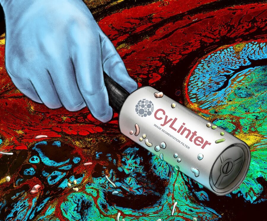
Baker, G. J., Novikov, E., Zhao, Z., Vallius, T., Davis, J. A., Lin, J.-R., Muhlich, J. L., Mittendorf, E. A., Santagata, S., Guerriero, J. L., & Sorger, P. K. (2024). Quality control for single-cell analysis of high-plex tissue profiles using CyLinter.
Nature Methods,
21(12), 2248–2259.
https://doi.org/10.1038/s41592-024-02328-0
Warchol, S., Troidl, J., Muhlich, J., Krueger, R., Hoffer, J., Lin, T., Beyer, J., Glassman, E., Sorger, P., & Pfister, H. (2024). psudo: Exploring Multi‐Channel Biomedical Image Data with Spatially and Perceptually Optimized Pseudocoloring.
Computer Graphics Forum, e15103.
https://doi.org/10.1111/cgf.15103
Nirmal, A. J., & Sorger, P. K. (2024). SCIMAP: A Python Toolkit for Integrated Spatial Analysis of Multiplexed Imaging Data.
Journal of Open Source Software,
9(97), 6604.
https://doi.org/10.21105/joss.06604
Wan, G., Maliga, Z., Yan, B., Vallius, T., Shi, Y., Khattab, S., Chang, C., Nirmal, A. J., Yu, K.-H., Liu, D., Lian, C. G., DeSimone, M. S., Sorger, P. K., & Semenov, Y. R. (2024). SpatialCells: automated profiling of tumor microenvironments with spatially resolved multiplexed single-cell data.
Briefings in Bioinformatics,
25(3), bbae189.
https://doi.org/10.1093/bib/bbae189
Nirmal, A. J., Yapp, C., Santagata, S., & Sorger, P. K. (2023).
Cell Spotter (CSPOT): A machine-learning approach to automated cell spotting and quantification of highly multiplexed tissue images [Preprint]. bioRxiv.
https://doi.org/10.1101/2023.11.15.567196
Muhlich, J. L., Chen, Y.-A., Yapp, C., Russell, D., Santagata, S., & Sorger, P. K. (2022). Stitching and registering highly multiplexed whole slide images of tissues and tumors using ASHLAR.
Bioinformatics, btac544.
https://doi.org/10.1093/bioinformatics/btac544
Rashid, R., Chen, Y.-A., Hoffer, J., Muhlich, J. L., Lin, J.-R., Krueger, R., Pfister, H., Mitchell, R., Santagata, S., & Sorger, P. K. (2022). Narrative online guides for the interpretation of digital-pathology images and tissue-atlas data.
Nature Biomedical Engineering,
6(5), 515–526.
https://doi.org/10.1038/s41551-021-00789-8
Schapiro, D., Yapp, C., Sokolov, A., Reynolds, S. M., Chen, Y.-A., Sudar, D., Xie, Y., Muhlich, J., Arias-Camison, R., Arena, S., Taylor, A. J., Nikolov, M., Tyler, M., Lin, J.-R., Burlingame, E. A., Human Tumor Atlas Network, Chang, Y. H., Farhi, S. L., Thorsson, V., … Sorger, P. K. (2022). MITI minimum information guidelines for highly multiplexed tissue images.
Nature Methods,
19(3), 262–267.
https://doi.org/10.1038/s41592-022-01415-4
Schapiro, D., Sokolov, A., Yapp, C., Chen, Y.-A., Muhlich, J. L., Hess, J., Creason, A. L., Nirmal, A. J., Baker, G. J., Nariya, M. K., Lin, J.-R., Maliga, Z., Jacobson, C. A., Hodgman, M. W., Ruokonen, J., Farhi, S. L., Abbondanza, D., McKinley, E. T., Persson, D., … Sorger, P. K. (2022). MCMICRO: a scalable, modular image-processing pipeline for multiplexed tissue imaging.
Nature Methods,
19(3), 311–315.
https://doi.org/10.1038/s41592-021-01308-y
Hoffer, J., Rashid, R., Muhlich, J. L., Chen, Y.-A., Russell, D. P. W., Ruokonen, J., Krueger, R., Pfister, H., Santagata, S., & Sorger, P. K. (2020). Minerva: a light-weight, narrative image browser for multiplexed tissue images.
Journal of Open Source Software,
5(54), 2579.
https://doi.org/10.21105/joss.02579
Goldberg, I. G., Allan, C., Burel, J.-M., Creager, D., Falconi, A., Hochheiser, H., Johnston, J., Mellen, J., Sorger, P. K., & Swedlow, J. R. (2005). The Open Microscopy Environment (OME) data model and XML file: open tools for informatics and quantitative analysis in biological imaging.
Genome Biology,
6(5), R47.
https://doi.org/10.1186/gb-2005-6-5-r47
Swedlow, J. R., Goldberg, I., Brauner, E., & Sorger, P. K. (2003). Informatics and quantitative analysis in biological imaging.
Science,
300(5616), 100–102.
https://doi.org/10.1126/science.1082602